MSK thoracic assessment

Warning

Other Concerns

Non MSK (e.g. visceral)

  • Rheumatology
  • Chronic pain
  • Osteoporosis
  • Fracture
  • Trauma

Other MSK

Anatomical and Biomechanical Considerations for Assessment of Thoracic Spine

There is great variability in the vertebral bodies within the thoracic spine. The upper thoracic vertebrae resemble cervical vertebrae and lower thoracic resemble lumbar vertebrae. In addition to this there are the additional articulations of the costovertebral and costotransverse joints with the ribs at all thoracic levels. Collectively the thoracic spine and ribs (as the chest wall) provide significant protection and stability. This is also enhanced by the thinness of the intervertebral discs.

The thoracic kyphosis is a primary curvature and its shape is determined by the vertebral bodies and discs. This kyphosis tends to increase with age but this change is also associated with reduced physical activity, poor postural habit, female gender and osteoporotic collapse. Increased kyphosis is generally asymptomatic.

The spinal canal is relatively narrow but the intervertebral foramen are relatively large. Therefore the incidence of radiculopathy in thoracic spine is much lower than in other areas of the spine. But, when present it is much more likely to be associated with symptoms of myelopathy. This creates a low incidence of radiculopathy but a higher rate of myelopathy in the presence of radiculopathy.

Spondylosis and bony degenerative changes are less evident within the upper and mid thoracic spine but a greater prevalence of Schmorl’s nodes (disc herniations within the central portion of the vertebral body) are seen on imaging but their clinical significance is unknown.

Movements of the thoracic spine are determined by direction and angle of the facet joints which varies from 0 - 90° in the different areas of the thoracic spine. But, overall segmental movement within the thoracic spine is much less than in the cervical or lumbar areas. Movement of the thoracic spine has rarely been investigated but it is cited that sagittal movements are greatest in the lower thoracic segments and rotation movements greatest in the upper thoracic segments. . With upper thoracic problems assessment of cervical spine movements need to be included and with lower thoracic spine problems, lumbar movements need to be included.

Assessment Aims

The main aims of the examination process are to:

  • Exclude serious spinal pathology
  • Assess the severity of the presenting complaint (in terms of pain, functional disability and psychological impact)
  • Formulate the diagnostic triage
  • Evaluate how and to what extent physiotherapy can be helpful


Diagnosis should be kept under review and not presumed definitive

Subjective Examination

The following information should be collected and documented through interviewing the patient:

  • Symptom site/description using body chart and numerical pain rating scale (NPRS)
  • Aggravating and easing factors
  • History of present condition (HPC)
  • Functional limitations
  • Progression or development of deformity with or without height loss
  • Social history (employment status/ nature of work/ hobbies/ smoking/ drinking/ activity levels)
  • Past medical history (PMH)
    • Including risk factors for osteoporosis
    • Previous history of cancer
    • Appropriate family history
  • Drug history (see also osteoporosis risk factors)
  • Previous treatment and outcomes/investigations and results
  • Assessment of patient’s fears, attitudes and beliefs in relation to their impairment
  • Identification of the patient’s agenda for consultation and their expectations of physiotherapy
  • Evaluation of the patient's understanding of the current problem

Red Flags

Thoracic specific conditions:

  • Discitis
  • TB/Potts disease
  • Cervical myelopathy
  • Cauda equina

Presence of serious pathology within the thoracic spine is less than 6%

Consider red flags and serious pathology guidance.

Yellow Flags

There is no specific literature concerning the presence of psychosocial factors (yellow flags) in patients with thoracic spine problems but there is significant evidence within the lumbar spine guidance and therefore awareness of these factors would appear to be important in all spinal conditions.

There is a general consensus that the most important predictor of determining outcome post an episode of musculoskeletal spinal pain is the patient’s psychosocial status. Linton (2000) concluded that psychological factors are strongly associated with the transition from acute to persistent pain and disability, and that psychosocial factors generally have a greater impact on disability than biomedical and biomechanical factors. Although the work by Linton does not specifically relate to thoracic pain it is possible that many of the factors identified will also affect recovery. The following psychological dispositions are consistent predictors of poor outcomes (Waddell, 1996):

  • A belief that pain is harmful or potentially severely disabling
  • Fear avoidance behaviour (avoiding a movement or activity due to a misplaced anticipation of pain) and reduced activity levels
  • Tendency to low mood and withdrawal from social interaction
  • Expectation of passive treatment(s) rather than a belief that active participation will help

It is therefore essential to screen for and document evidence of psychosocial barriers to recovery (yellow flags). These provide indicators on the risk of chronicity developing. An assessment of the psychosocial status of the patient should be made with regard to the following (Kendall et al., 2004):

A = Attitudes and beliefs about pain

B = Health behaviours

C = Compensation issues

D = Diagnoses and treatment issues

E = Emotions

F = Family

W = Work related issues (see blue and black flags)

If appropriate, screen for blue flags. Blue flags are individually perceived occupational factors that impede recovery and return to work (RTW) (Kendall et al., 2004). Examples include low job satisfaction or inadequate support from supervisor.

Identify black flags. Black flags are actual (organisational) barriers to RTW. These relate to social security and healthcare systems (Kendall et al. 2004). Examples may include adverse sickness policies or benefit systems.

A person is at risk of developing disability if there is a cluster of a few salient yellow flags or a group of less important yellow flags that combine cumulatively.

Objective Examination

The objective assessment should be tailored to the individual patient and their subjective history.

If an identifiable cause of thoracic pain is found, then the examination and treatment should be tailored accordingly. Evidence within these guidelines for the reliability and validity for the majority of diagnostic tests used by therapists is limited. Diagnosis is hence made on the basis of theoretical knowledge and sound clinical reasoning.

The objective examination should focus on the patient’s functional abilities, presence of structural deformity, cultural sensitivities and their willingness to participate as opposed to finding a physical cause of their thoracic pain or radiculopathy.

Assessment

May include:

  • Examination of posture including height loss
  • Body composition (BMI, body habitus, breast size)
  • Identification of structural deformities
  • Neurological examination (predominantly altered sensation)
  • Range of movement/functional movements (including cervical and lumbar spine movements as required)
  • Assessment of neural mobility
  • Palpation
  • Repeated movement testing
  • Gait

Neurological examination should always be undertaken whenever signs or symptoms are of an atypical pattern/presentation, where non-musculoskeletal pathology is suspected or where neurological pathology is suspected which may include the presence of upper and/or lower limb symptoms.

This neurological examination should include:

  • Upper and lower limb dermatomes
  • Upper limb myotomes (myotomal weakness should be recorded in relation to the Medical Research Council (MRC) grading system for muscle power (0 – 5)
  • Upper and lower limb reflexes (biceps, brachioradialis, triceps, knee, ankle)
  • Hoffman’s sign
  • Lower limb reflexes
  • Babinski
  • Clonus

Wikimedia commons. Depiction of dermatomes of the human body.

Visual depiction of dermatomes of female body


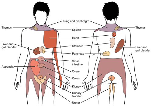
WikiMSK. Visceral pain.

Depiction of visceral sensations map of the body

Analysis

On completion of the assessment the therapist should be able to:

  • Confirm the diagnostic triage
  • Formulate a clinical impression that attempts to identify the dominant cause of the patient’s pain/symptoms i.e. are the symptoms predominantly from the muscular, articular or nervous system. There is some evidence to support the reliability of diagnosis with a sub-classification system e.g. McKenzie Mechanical Diagnosis and Therapy. Generating a clinical impression facilitates the clinical reasoning process (NICE, 2015)
  • Where possible identify the dominant pain mechanism (nociceptive, peripheral neuropathic, central or sympathetic)
  • Summarise the patient’s main complaints in terms of physical, psychological and social factors
  • Negotiate agreed goals and formulate a time-directed treatment plan
  • Identify and record appropriate outcome measures
  • Consider input from other services

Outcome Measures

Standard 6 of the CSP Standards of Professional Practice (2005) states that it is an explicit requirement for members to use published/validated outcome measures in routine clinical practice. The outcomes should be clearly identified and documented at the beginning and end of treatment and at the end of the episode of care. Validated outcomes commonly used include:


It is generally acknowledged that treatment outcomes should be functionally driven and goal orientated. The GG+C NPRS and patient specific functional scale conforms to both of these criteria.

At the end of each treatment episode, a statement or discharge summary should be drawn that clearly states the reasons for concluding treatment and highlights the outcome of the episode of care. This should reflect on:

  • The diagnostic triage
  • Interventions received
  • Functional outcome. This may include the patient’s understanding of their problem and their role (plus that of the GP) in their ongoing management.

Where referral to another professional is indicated, clear reasons for this should be made to the patient and documented.

Evidence

Kendall, NAS, Linton, SJ, Main CJ (1997). Guide to assessing Psycho-social Yellow flags in Acute Low back pain: Risk factors for long-term disability and work loss. Accident Compensation Corporation and the New Zealand Guidelines Group, Wellington, New Zealand. 2004 edition.

Linton, SJ. Psychological factors in neck and low back pain in: Nachemson, AN and Jonsson, E (eds) Neck and Back Pain: The scientific evidence of causes, diagnosis and treatment 2000 Lippincott Williams and Wilkins, New York.

Nice – Non Specific Neck Pain: https://cks.nice.org.uk/neck-pain-non-specific

Waddell G, Feder G, McIntosh A, Lewis M and Hutchinson A. Low Back Pain Evidence Review. Royal College of General Practitioners. London: 1996

Editorial Information

Last reviewed: 30/04/2024

Next review date: 30/04/2025

Reviewer name(s): Louise Ross, Alison Baird, Karen Glass.