Warning

Objectives

Epidural analgesia

Audience

  • Argyll & Bute HSCP and Highland HSCP
  • Secondary Care

Please note this guidance is for use in in-patients and is not designed for managing patients with chronic pain

  • Epidural infusions for acute pain may be managed in the following areas:
    1.  Recovery, Main Theatres
    2.  ITU
    3. Surgical High Dependency
  • The responsibility for ensuring safe and effective epidural analgesia after surgery rests with the Anaesthetist that instituted the epidural and with the Department of Anaesthesia. Changes to the epidural prescriptions may only be authorised by an Anaesthetist or Acute Pain Nurse
  • Epidural infusions are delivered by a dedicated Epidural pump only.
  • Insertion of epidural catheter is an aseptic technique, performed in the anaesthetic room in theatre. The catheter can remain in-situ for a maximum of 96 hours. If epidural analgesia is required for longer, the decision must be agreed by the anaesthetist who inserted the epidural or the ITU consultant anaesthetist.
  • The Acute Pain Team (bleep 1003 or 6056) or in their absence, the ITU anaesthetist should be contacted for any of the following epidural problems: inadequate pain relief; motor block; hypotension that has not improved with treatment; high sensory block; signs of local anaesthetic toxicity; patient sedation or confusion.
  • Levobupivicaine 0.125% bags must be stored in a locked cupboard away from other intravenous fluids/antibiotics.

Setting up an epidural infusion

Can be completed by any of the following who have received training in the use of the epidural pump: Anaesthetists; Anaesthetist assistants; Acute Pain Nurses

  • Attach the infusion bag to the epidural giving set. Attach drug label to the bag.
  • Programme the Epidural pump and purge the line according to the manufacturer’s instruction and as per training.
  • Lock the pump using the digital access code.
  • On arrival in recovery, two recovery nurses must check the pump programme against the prescription sheet to ensure that it is correct.
  • Prior to discharge from recovery the pump and the prescription must be checked. Ensure yellow sticker (PCA/epidural opiates is in progress) is on the front of the Kardex.

Procedure

  • Epidural catheters must be inserted using full aseptic technique and should preferably be sited in theatres or ITU.
  • The Acute Pain Service can advise on the epidural fixation device to be used. The use of glue as a fixation technique is recommended by the Acute Pain Service. Please contact the Acute Pain Team for further information.
  • The epidural filter should be secured to the upper arm or chest wall with a padded dressing, preventing the catheter from being pulled out of the connector. If the line does become disconnected at this point (i.e. distal to the filter) the epidural catheter is inevitably contaminated and must be removed.
  • Epidural bags with fentanyl must be renewed every 24 hours.
  • The skin exit site should be inspected every nursing shift. If there are any signs of infection (tenderness, inflammation or exudation), the Acute Pain Nurse or ITU Anaesthetist should be informed. If the epidural is removed, the tip should be sent to bacteriology for culturing.
  • After 96 hours the giving set and filter should be changed

Preparation or change of epidural infusion (is the flow chart to be deleted or replaced?)

Two nurses to be present when preparing an epidural infusion 

  • Epidural prescription sheet
    • 0.1 % levobupivacaine with fentanyl 2 microgram/mL
  • Levobupivicaine bag with fentanyl 2 microgram/mL to be checked by 2 nurses from controlled drugs cupboard according the hospital policy: Preparation and administration of controlled drugs (see resources)
  • Check levobupivicaine bag for any puncture, discolouration, haziness and crystalline or other particles. Check expiry date
  • Check the patient identity against the prescription chart. Check that the epidural pump has been programmed correctly and corresponds to the epidural prescription sheet.
  • Complete the controlled drug register according to hospital policy. Sign commencement of epidural in comments section on prescription sheet.

(Please contact acute pain nurse bleep 1003 if further supervised practice is required).

Epidural nursing management and monitoring (adult)

Nursing Management

Patients with epidural infusions may only be nursed on designated wards and be cared for by registered nurses who:

  • hold a valid (3 yearly) certificate of attendance of the Acute Pain Service epidural lecture,
  • declares her/himself to be competent,
  • received epidural pump training by ward pump trainer, acute pain nurse or on-line pump training video.

Oxygen 4 L/min via Hudson mask or 2 L/min via nasal cannula should be given to patients for 48 hours post-operatively and then overnight until the epidural infusion is discontinued, unless prescribed otherwise by the anaesthetist.

Unless authorised by a senior anaesthetist, no other opioids should be given by any other route, whilst epidural infusion is in progress.

Naloxone injection should be readily available on all wards were epidural infusions are managed and registered nurses should know where it is located.

All patients who have an epidural infusion should have a patent IV cannula in case of adverse reactions.

Monitoring

1. Blood pressure, Pulse, Respiratory rate, Oxygen Saturations and Sedation score should be recorded on the NEWS chart:

  • In recovery: every 15 mins
  • On ward: 
    • 1/2 hourly for first 1 hour
    • Hourly for 4 hours
    • 2 hourly for 12 hours
    • Then 4 hourly until 24 hours after stopping infusion

If a patient receives a “top-up” of levobupivacaine stronger than 0.125%, the patient must have their BP recorded every 5 minutes for 30 minutes.

2. Motor score must be recorded on the epidural chart:

  • In recovery: every 30 mins
  • On ward:
    • 1/2 hourly for 1 hour
    • Hourly for 4 hours
    • 2 hourly for 12 hours
    • Then 4 hourly until 48 hours after stopping the infusion

3. Sensory level is recorded on the epidural chart. Use ice to detect level.

  • In recovery: on anaesthetist’s instruction and before leaving recovery.
  • On ward: at every change of shift and if any of the following are noted:
    • inadequate pain relief
    • increased motor block

Also remember to check sensory level if epidural infusion has been stopped for any reason, before re-starting and one hour after
increasing rate.

4. Nausea score is to be recorded on the NEWS chart.

  • In recovery: every 15 mins
    • On ward: 1/2 hourly for 1 hour
      Hourly for 4 hours
      2 hourly for 12 hours
      Then 4 hourly thereafter.

5. Pain score is to be recorded on the NEWS chart.

  • In recovery: every 30 mins
    • On ward: 1/2 hourly for 1 hour
    • Hourly for 4 hours
    • 2 hourly for 12 hours
    • Then 4 hourly thereafter.

6. Epidural Exit Site: While the epidural infusion is in progress, the exit must be checked on every shift for signs of leakage or infection. Once the epidural catheter has been removed, the exit site must be checked daily until the patient is discharged from hospital. This must be documented on the NEWS chart. Any signs of infection must be reported to the Acute Pain Team or ITU anaesthetist.

7. Temperature must be recorded on the NEWS chart every 4 hours (or more frequently, if the patients condition requires it)

8.Pump recordings must be recorded on the epidural chart as per Hospital policy, ensuring that the 'amount delivered' and the 'amount remaining' add up to the original starting total.

Inadequate analgesia from epidural infusion (adult)

If the patient’s pain score is higher than 4 for more than one set of observations, follow below.
Firstly assess the location of the patient’s pain.

If patient has received a “top-up” of levobupivacaine stronger than 0.125%, the following observations should be carried out.

  • Record blood pressure every 5 minutes for 30 minutes.
  • If required, the patient can then be transferred out of bed 45 minutes after “top-up” if BP stable.
  • If “top up” given in recovery and BP is stable then patient can be transferred to the ward after 30 minutes.

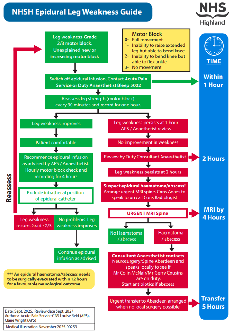
Epidural leg weakness

Measuring sensory level, motor block and patient controlled epidural analgesia

Measuring Sensory Level

This helps us identify which nerves have been blocked by the local anaesthetic. We assess thermoreceptors at a dermatomal level using a cold stimulus (such as ice) only. It is useful to have a supply of ice cubes in the freezer compartment of your fridge. The ice cube can be placed in either a disposable glove and in a gauze for single patient use.
How to perform the sensory level measurement:

  • Explain the procedure to the patient.
  • Place the ice cube on a part of the patient’s body that is not affected by the epidural e.g. the patient’s forehead and ask the patient if it feels cold.
  • Next, place the ice cube below the level of the wound area and work upwards, placing the ice cube at each dermatomal level and on both sides of the body, thus checking that the block is bilateral. (This can also be done on the patients back).
  • When the patient feels the ice as cold (the same as when placed on their forehead) it is usually a good indication of how far the block has spread. Using the body chart on the epidural laminated guidelines for reference, you will be able to determine the dermatomal level.
  • If the patient feels the cold stimulus all over the area that is being tested and the patient’s pain is controlled do not be alarmed. This occasionally occurs when weak local anaesthetic solutions are used which do not have an effect on the thermoreceptors.

Measuring Motor Block

Epidural infusions used for post-operative pain relief should not cause the patients legs to become weak. The motor nerves are not normally affected by the weak solution of local anaesthetic used.

This is a side effect that must be acted upon and treated.

  • All patients must have their motor block score performed and recorded at the same time as their vital sign observations.
  •  If the motor block is >0, the epidural must be temporarily stopped and the Acute Pain Nurse or the ITU Anaesthetist contacted.

Motor Block Score

0 = Full movement
1 = Inability to raise extended leg but able to bend knee
2 = Inability to bend knee but able to flex ankle
3 = No movement

Patient Controlled Epidural Analgesia (PCEA)

  • All PCEA must be prescribed on the epidural chart.
  • Only patients who have had major surgery, who are mentally alert and understand the concept of PCEA should be considered for its use.
  •  If the epidural catheter is inserted at a level corresponding to the upper third of the inclusion the following setting are suggested:
    Infusion rate = 0mL to 10mL per hour
    PCEA setting = 5mL PCEA bolus
    Lockout = 20 minutes (3 bolus in an hour)
  • All epidural observations should be carried out as per the standard monitoring requirement.

Epidural complications

Respiratory Depression

If the respiratory rate is less than 9 breaths/per minute (moderate)
  1. Stop the infusion and administer oxygen 10L per minute via a facemask if 02 sats <95% Ensure a clear airway.
  2. Call the Acute Pain Nurse or ITU Anaesthetist, as it may indicate that the opioid needs to be reduced or removed from the infusion.
  3. Monitor sedation score, respiratory rate and oxygen saturation every 15 minutes, until the respiratory rate is greater than 10 per minute.
If the respiratory rate is less than 7 breaths/per minute (severe)
  1. Stop the infusion and give oxygen at 10L per minute via a facemask.
  2. Call the Ward Doctor or Nurse Practitioner immediately.
  3. Call the Acute Pain Nurse or ITU Anaesthetist.
  4. Administer NALOXONE. See Naloxone guidance

If the patient is apnoeic, call the arrest team and initiate CPR.

Sedation

If the sedation score is 2: treat as for moderate respiratory depression.
If the sedation score is 3: treat with naloxone as for severe respiratory depression.

Sensory Level

  • If the sensory level is higher than stated on the epidural prescription sheet and/or the patient is showing signs of a high block (e.g. difficulty breathing, numbness in hands or arms) stop the infusion for 30 minutes and sit the patient up as tolerated. Recommence the infusion at half the rate of the previous infusion rate. Re-assess the sensory level after 30 minutes.
  • If the patient has a high sensory level and increasing motor block, stop the infusion and contact the acute pain nurse or ITU anaesthetist.
  • If the patient is on PCEA and the block is high, stop the infusion and contact the Acute Pain Nurse or ITU anaesthetist.

Hypotension

If the blood pressure is less than that stated on the front of the epidural chart or if it is significantly less than the pre-operative value, carry out the following :

  • Lie the patient flat with one pillow under the head and with the legs raised slightly. Do not tip head down. 
  • Stop the pump
  • Give oxygen at 10L per minute via facemask if saturations higher than 95%. Contact the ward doctor or nurse practitioner.
  • Check the sensory and motor block levels to assess if the epidural block is too high. If it is considered that the epidural infusion is causing the hypotension, or if the BP has not improved despite appropriate management, contact the ITU anaesthetist. When the BP is satisfactory, restart the pump.

Confusion and Hallucinations

  • Check that there is no other underlying cause e.g. hypoxia, infection.
  • Contact the ward doctor if any signs of above.
  • Once all the other factors have been excluded, if patient is elderly and/or has reduced renal function contact the Acute Pain Nurse or ITU Anaesthetist who may consider reducing or removing the opioid from the epidural.

Urinary Retention

Most patients will have a urinary catheter in situ. If the urine output drops, check that the urinary catheter is not blocked as patients may not complain of a full and painful bladder.

Pressure Area Care

Patients with an epidural infusion may have reduced sensation. All patients must be scored as having a high risk of pressure sores.

Motor Block

  • The patient may have a motor block score of 2 or 3 on return from theatre but this should regress to 0 or 1 within 4 hours.
  • If the motor block score does not improve or increase to 2 or 3 after initial recovery, or at any time during infusion, stop the pump and contact the Acute Pain Nurse or the ITU Anaesthetist.
  • Refer to treatment of epidural complications guideline.

Local Anaesthetic Toxicity 

Early signs and symptoms Late signs and symptoms 
tinitus profound hypotension 
flushed face bradycardia, ventricular arrhythmias 
circum-oral numbness toni-clonic convulsions 
lightheaded drowsiness 
slurred speech coma 
hypotension respiratory arrest 
muscle twitching cardiac arrest - this is compounded by hypoxia
If any of the signs and symptoms of local anaesthestic toxicity are present:
  • Stop infusion
  • Contact the ITU Anaethetist urgently
  • Administer Oxygen 10L per min via facemask if O2 Sats < than 95%.
  • Call for help and initiate CPR procedures if the patient is apnoeic. Call 2222.
  • Obtain Lipid rescue bags: ClinOleic 20%, stored in pharmacy cupboard, surgical HDU, theatre corridor (bleep 1089 for access) and labour suite and commence as soon as possible.

Removal of epidural catheter

Registered nurses who have completed the acute pain service epidural training and have maintained their skills may remove an epidural catheter. If further supervision required contact acute pain nurse (bleep 1003 or 6056).

Timing of Removal

Epidural catheters should preferably be removed in the morning so that the patient’s neurological condition can be observed.

  • Any complaint of new back pain, increased motor block and difficulty in passing urine or any change in neurological function should be reported to the Acute Pain Team or the ITU anaesthetist immediately.
  • Low Molecular Weight Heparin (e.g. Clexane). Epidural catheters must not be removed until 12 hours have elapsed after the last dose. The subsequent dose of low molecular weight heparin must not be given for a further 4 hours after removal.
  • Standard or unfractionated heparin given on a twice daily basis. Epidural catheters should not be removed until 8 hours after the last dose of heparin. Further doses of heparin should not be given for at least 2 hours after removal.
  • Patients on continuous heparin infusions need advice from the anaesthetist and surgeon responsible for the patient.

High risk coagulation patients. I.e. liver resection

  • Ensure high risk sticker is attached to dressing on epidural exit site.
  • Ensure COAG screen is taken on the morning of the day the epidural catheter is to be removed.
  • Inform the acute pain nurse or ITU anaesthetist of the results and they will confirm if epidural catheter can be removed.

Disposal of Medicines

Please dispose of levobupivicaine or levobupivicaine and fentanyl bags in appropriate medicines container (large sharps bin with blue lid).

  1. Procedure
    • Gather relevant equipment required:
    • trolley
    • dressing pack
    • skin disinfectant e.g. chlorhexidine gluconate
    • airstrip plaster (also required, if tip to be sent to bacteriologyuniversal container
    • sterile scissors
  2. Explain the procedure to the patient. The patient can choose between one of the two positions: lying on their side, with knees drawn up slightly or sitting upright, bent forward over a pillow.
  3. An aseptic technique must be used throughout the procedure. Wash hands thoroughly before starting the procedure. Remove the dressing and wash hands again.
  4. Clean the area around the catheter insertion site. Place a sterile swab on the skin at the catheter insertion site. With another sterile swab, hold the epidural catheter and gently pull the catheter to remove it. The resistance is normally slightly stronger than when removing an IV cannula. If the resistance is more than this, stop the procedure and call the Acute Pain Nurse or Anaesthetist for assistance.
  5. Once the catheter has been removed, cover the site with the plaster and settle the patient into a comfortable position.
  6. Check the blue tip is present and intact and record on prescription sheet.
  7. If there are any signs of infection or discharge, a sterile dressing should be applied. Do not use an occlusive type dressing. Also inform the Acute Pain Nurse or ITU Anaesthetist.
  8. The tip of the epidural catheter should be sent to Microbiology for Culture and Sensitivity if:
    • There are any signs of infection at the exit site
    • The patient has an unexplained temperature over 38 degrees centigrade
    • The doctor requests it as part of their investigation
    • The epidural catheter has been in situ for more than 96 hours.
      Document on the epidural chart if the tip has been sent to bacteriology.
      If the epidural catheter has fallen out it will not be suitable for culture and sensitivity.
  9. Motor score monitoring should be recorded for 48 hours after infusion stopped. The epidural exit site should be inspected every day until discharge.  This should be noted on the NEWS chart in the spare monitoring column.

Editorial Information

Last reviewed: 31/10/2022

Next review date: 31/10/2025

Author(s): Acute Pain Team.

Version: 3

Approved By: TAMSG of the ADTC

Reviewer name(s): Louise Reid, Clinical Nurse Specialist, Claire Wright Acute Pain Nurse Specialist .

Document Id: TAM110

Related resources

Further information for Health Care Professionals

  • Controlled drugs policies and SOPs via the Policy Library (see under PPGs ratified documents (NHS HIghland Intranet access required)
References

Self-management information