Obstetric Anaesthesia Handbook

Introduction

The initial version of this handbook was produced in 2002 and based on work done by Dr. Justine Nanson, Ninewells Hospital, Dundee, to whom we are indebted. Since then it has undergone several revisions and updates. It is intended to facilitate learning and provide guidance for management of patients under your care. It is not an alternative to contacting a more experienced obstetric anaesthetist in cases of doubt.

The handbook is available in the printed form (“seminar”/anaesthetic pigeonhole room within main theatres, and in the anaesthetic room of the labour suite theatre). If you would like extra paper copies, please ask Shelby Noble, the anaesthetic secretary. If you would like the handbook to be emailed to you, please ask Dr Mairi Crawford (Mairi.Crawford@ nhslothian.scot.nhs.uk). A copy is also on the shared drive and on the Lothian Clinical Companion app.  

We like to know if unusual or challenging events occur. If there is an unusually sick patient or you come across something outside of your experience, please try to contact one of us. Any questions, comments or suggestions, please do not hesitate to contact us.

This version of the handbook has been modified for St John's Hospital. Please contact Dr Mairi Crawford if you notice any mistakes/ discrepancies in it. (Email: Mairi.Crawford@ nhslothian.scot.nhs.uk).  

Please also be aware that certain protocols are in the process of being updated. We will try to keep the most up to date version of the handbook in hard copy form in theatres.


The obstetric anaesthetists who have regular obstetric sessions at St John’s Hospital are currently:


Mairi Crawford (lead clinician)

Richard Burnett                     Claire Caesar              Lynn Carragher

Sarah Cross                            David Falzon              Clare Gardner

Nicola Hogan                         Audrey Jeffrey           Bridget Podmore

Rachel Smith                          Patrick Ward              Karen Watson


Other consultants also have an interest in obstetrics and cover sessions on a less regular basis.


2022 Update

The handbook has been brought up to date following review and departmental consensus, and trainee feedback. In addition to review of the overall contents, features have been added to improve the utilisation of the handbook as a dual online/hard-copy resource. A collection of “quick reference” resources have been collated at the front. We hope you find this resource useful during your attachment.

Daily Duties of the Obstetric Anaesthetist

We expect that anaesthetists working in labour ward consider themselves part of the team involved in intrapartum care. You should maintain regular communication with the midwives and obstetricians in order to anticipate problems and avoid crises.

On arrival at 08.00 take a handover from the anaesthetist who has been on-call. You need to know how many patients are in labour ward (with or without epidurals), what admissions are expected and about any problems that might be anticipated.

There is a daily handover meeting with obstetricians/midwives/anaesthetists at 08.30 in labour ward, where the previous 24 hours’ deliveries are discussed, as well as current labour ward patients.

Confirm clinical priorities; see and assess patients for theatre.

Discuss potential problems and the plan for the day with the consultant anaesthetist.

Check the anaesthetic machines in the labour ward theatre. Check location of trolleys with equipment for difficult/failed intubation, defibrillator and massive haemorrhage.

Check that there are fresh drugs for emergency Caesarean section and management of acute hypotension following regional block in the fridge.

GA drugs:

Thiopentone 500 mg (pre-prepared and supplied from pharmacy)

Suxamethonium 2x 100mg (with 5mL syringe)

Atracurium 50 mg (ampoule left unopened in tray with syringes/needle/label).

Other emergency drugs:

Atropine 600 mcg

Phenylephrine 20 mcg/mL - (10mg into 500mL bag of normal saline)

Ephedrine 30 mg

Syntocinon 10iu


Be available promptly by page 3948 for patients requesting epidural analgesia, patients who require operative intervention or resuscitation.

Ensure you complete relevant paperwork for each clinical task you perform. Fill in a blue audit chart when you do any anaesthetic intervention, and then put it into the box for completed audit forms in the anaesthetic room in the labour ward. Remember to fill in any additional interventions you perform on the same patient eg, resite epidural, caesarean section etc. Complete any additional audit forms as required (eg ERAS, WardWatcher).

Carry out follow-up visits at weekends (usually done by consultant during week).

Useful Contact Numbers


To bleep dial 110 on any internal hospital extension, the bleep number, followed by the extension from which you are bleeping.

Anaesthetist ICU Bleep 3561
Anaesthetist (Obstetrics) Bleep 3948
Consultant Obstetric Anaesthetist (8-5 weekdays) Bleep 3765

Obstetric Consultant (8-5 weekdays)

Bleep 3898

Obstetric Reg

Bleep 3558

Obstetric SHO (8-5 weekdays)

Bleep 3578
Obstetric SHO (out of hours) Bleep 3559
Theatre Coordinator Bleep 3541
ODP on-call Bleep 3656

Blood transfusion

Dial 53354

Haematologist on-call (lab, not medic)

Bleep 3729
Biochemist on-call Bleep 3728

SCBU/NICU

Dial 54394
Neonatal Paeds Reg Bleep 3565

Analgesia in Labour

Assessment of the parturient requesting epidural analgesia

Epidural analgesia should be considered for all labouring women in the labour suite who request it. However, epidural analgesia is not without potential complications, so it is essential to assess patient suitability. Assessment should aim to identify or exclude potential contraindications and ensure that it is safe to proceed.

First ensure that the patient wishes epidural analgesia and obtain informed verbal consent. Ensure this is witnessed by the midwife or birthing partner, and documented.

Ask what the patient knows about epidural anaesthesia and discuss hypotension, headache, backache, neurological damage, failure of analgesia and subsequent treatment options as appropriate, and answer any questions patient has. Explanation to the mother (and her partner, if available) must be in terms appropriate to the situation. There is no point in giving too comprehensive an explanation to a mother who is contracting painfully every two minutes if she is unable to give meaningful consent to anything! 

Obtain a history (from patient, notes or midwife) of:

  • cardiovascular or respiratory disease, which may indicate limited ability to cope with epidural complications
  • neurological/back problems which may cause concern about epidural placement and post-procedure outcome
  • coagulation problems
  • drug history - especially anticoagulants, allergy
  • previous experience of local/general anaesthesia
  • pregnancy problems e.g. pregnancy induced hypertension, antepartum haemorrhage (APH).

Many of these problems will have been identified during antenatal care and triggered referral to the Obstetric Anaesthesia clinic – check Trak for an Anaesthetic Management Plan.

Examination

  • check vital signs - pulse & blood pressure
  • temperature, if suspect systemic infection
  • assess the patient’s airway – in all GA will be the back-up plan in event of failed regional anaesthesia
  • check insertion site to exclude infection & assess anatomical difficulty
  • ask the midwife if there are concerns about fetal wellbeing (CTG changes, fetal scalp pH).
Indications for and contraindications to epidural insertion

Pain relief in labour may be provided by non-pharmacological means, or by entonox, opioids or regional techniques

There are two main indications for epidural analgesia in labour:

Group 1: Maternal request for analgesia in spontaneous labour.

It must be appreciated that provision of epidural analgesia for normal uncomplicated labour is a medical intervention, with attendant risks and side-effects. As long as this is understood and accepted by the mother, epidural analgesia should be made available at her request unless medically contraindicated. The time from informing the anaesthetist of the mother’s request until attending the woman should not ideally exceed 30 minutes. It should definitely not exceed 60 minutes, other than in exceptional circumstances.  If necessary, due to volume of other emergencies out of hours, you should contact the on-call consultant, who may be able to help.

Group 2: Obstetric/medical indications for epidural analgesia. (Groups, which fall into the category of complicated (or potentially complicated) labour.
  • multiple pregnancy
  • malpresentation (occipito-posterior position, breech)
  • intrauterine growth restriction
  • pregnancy induced hypertension/pre-eclampsia
  • slow progress in labour
  • previous caesarean section (VBAC)
  • diabetes (gestational or pre-existing)
  • uncoordinated uterine activity managed by oxytocin.

These mothers should be counselled regarding epidural analgesia, in view of their increased chance of needing instrumental or operative delivery. The aim is to avoid the need for emergency general anaesthesia with its attendant risks.  Many of these patients will have attended the Obstetric Anaesthesia clinic, and will have been counselled on the benefits of early epidural in labour. As above, the Anaesthetic Management section of Maternity Trak should provide a suggested plan.

Contraindications to epidural insertion:
  • patient refusal
  • sepsis (local or generalised)
  • hypovolaemia/shock
  • raised intra-cranial pressure
  • abnormal coagulation (pathological or iatrogenic).

If in doubt, discuss with a senior colleague. Ultimately this should be a shared decision between the woman, anaesthetist, and maternity team, and the discussion about risk individualised as far as possible.

Insertion of an epidural catheter for obstetric analgesia/anaesthesia
Preparation of patient

Ensure suitability and record baseline vital signs.

Explain technique to patient. Insert at least a 16g intravenous (IV) cannula, preferably in the non-dominant forearm/hand, using local anaesthesia. Ask midwife to assist.

Use an aseptic technique

Ensure back sprayed with 0.5% Chlorhexidine spray before opening epidural pack (either yourself or the midwife to spray, 4-6 sprays required). Scrub hands and arms; wear protective/aseptic clothing (hat, mask, gown & gloves)

Prepare equipment aseptically (swabs, 18G or 16G epidural kit, sterile saline, clear occlusive dressing +/- fixation device, local anaesthetic solution to test and/or initiate block)

The procedure (mid-line approach)
  • Place patient in preferred position (sitting/lateral) Inform patient of all further manoeuvres before/as they happen.
  • Ensure that the Chlorhexidine has had time to dry. Place sterile drape to cover back & hips.
  • Palpate the hips to establish position of interspaces (Tuffier’s line, drawn between the iliac crests, usually, but not always, crosses the L3/4 interspace. It lies slightly higher in the pregnant state, because of pelvic rotation). Using this as a guide select a suitable space for epidural insertion – usually L2/3, L3/4 or L4/5.
  • Use lidocaine 1 or 2% to anaesthetise area around insertion site. A needle (e.g. 19 G) can be used to pierce the skin.
  • Insert epidural needle slowly & carefully through skin, supra-spinous and interspinous ligaments until held in place by ligaments. Remove obturator, and attach loss of resistance syringe containing saline. Place hand holding needle against patient's back to avoid sudden needle movement. Advance needle with constant pressure on syringe plunger until loss of resistance is felt.
  • If no leak of blood/CSF through needle is seen, insert catheter into epidural space. Carefully remove needle over catheter. Never attempt to withdraw the catheter through the needle, as the catheter may break.
  • Withdraw the catheter so that there is enough left within the epidural space to prevent dislodgement – usually 4 - 5cm left in space, and greater in morbidly obese patients.
  • Attach adapter. Aspirate using a 2 ml syringe to ensure no blood/CSF.
  • Prime filter with local anaesthetic solution and attach to the adapter.
  • Cover the insertion site with the clear dressing. Ask an assistant to fix the edges of the dressing and the catheter to the skin with suitable tape.
  • Inject test solution, asking the patient to report tinnitus, paraesthesia, palpitations, or leg weakness. Assess Bromage score before commencing epidural infusion.
  • Document procedure on an anaesthetic chart & audit form.
Initiating epidural analgesia

Once an epidural catheter has been inserted then analgesia must be started. How this is achieved depends to a certain extent, on the planned maintenance regime (see p 10) but it must be ensured that no harm will result if the catheter should migrate into a vein or the CSF.

A test dose (4ml of 2% lidocaine) may be given to detect intravascular or intravenous placement. All doses of local anaesthetic, particularly the first one, should ideally be fractionated (e.g. 5ml of solution, at least 5 minutes apart and over at least 1 minute), although in an emergency this may be modified.

Required level of block:

            1st stage:                    T10 to L1

            2nd stage:                    T10 to S3

Potential complications include local anaesthetic/opioid toxicity or high spinal anaesthesia.

Initiate analgesia with the following:

  • 15ml of the “bag mix” (i.e. 0.1% levobupivacaine + fentanyl 2mcg/ml): give slowly. Assess analgesia after 15 minutes.
  • Check maternal pulse and blood pressure every 5 minutes for 20 minutes after every dose.

Maintenance of epidural analgesia

Continuous infusion

Maintenance of anaesthesia is generally provided by a constant infusion of pre-mixed solution at 10ml/hr via a dedicated epidural infusion pump. Midwives will record hourly observations. A certain percentage of patients receiving this technique will require a 'top-up' at some point. Many midwives at St John’s have completed a training programme and are permitted to give top ups of the ‘bag solution’ providing you prescribe them on the appropriate place on the anaesthetic record. Usual midwife top-ups would be 10 mls of the bag solution (levobupivacaine 0.1% with fentanyl 2mcg/ml), every 30 minutes.    

PCEA and PIEB

An alternative to providing a continuous infusion of bag mix is to use a combination of patient-controlled epidural analgesia (PCEA) with programmed intermittent epidural boluses (PIEB). The programmed component can deliver a bolus of between 5 and 10mL bag mix every hour, with a PCA bolus of 5-10mL on a 15 minute lockout.

Evidence suggests that this provides greater sensory dermatomal spread with reduced motor blockade. This is dependent on new epidural pump technology which is being introduced at St. John’s in mid-late 2022. There is a training module on the company website for programming the pumps and the protocol is yet to be finalised.

Management of the second stage

There is no clear evidence that allowing the epidural to wear off in the second stage of labour increases the chance of spontaneous vaginal delivery. It is inhumane to provide analgesia throughout the first stage of labour and then allow the mother to experience the severe pain of the second stage. Moreover, if the epidural has been allowed to wear off in the second stage, and instrumental or operative delivery becomes necessary, there may be insufficient time to extend the block.

Management of an inadequate block

Approximately 10% of women will have initially unsatisfactory blocks, and around 2% of these will be persistently inadequate. Blocks should be carefully monitored so that inadequacies can be detected early. If asked to assess an inadequate block, the first thing to do is check the position of the catheter: the commonest reason for a previously satisfactory block failing is the epidural catheter falling out. Test and record the pattern of sensory/motor block. Pump malfunction is another cause to look for.

Limited unilateral spread

Block is usually confined to the lumbar dermatomes closest to the insertion site of the catheter; this is due to the catheter passing out from the epidural space, into the intervertebral foramina.

Unilateral block

Distribution of solution to only one side of the epidural space may be due to a dorsal midline septum arising from the posterior aspect of the dura mater, acting as a barrier to the free spread of local anaesthetic solution. It is more common in patients with scoliosis.

Missed segments

Less common than above, may also be related to isolation of nerve roots by longitudinal septae.

Inadequate spread

If cranial or caudal spread is insufficient despite top-ups, it may be due to horizontal septae. It is more common in patients who have had previous spinal surgery, presumably due to scarring in the epidural space.

Management options

The following manoeuvres can be tried:

  • pulling back catheter if > 3 cm into space
  • lying mother with unaffected side down and giving further top-up
  • increase the concentration of top-up solution if previously the “bag” solution top-ups are not sufficiently effective.
  • opioid top-up (50 mg fentanyl)
  • A top-up with 50-100mg clonidine may be considered (repeated 8 hourly)

Re-siting the catheter early is often a better option than repeated attempts at adjustment.

A paramedian approach may help if re-siting the catheter, as catheters inserted this way travel a straighter course in the epidural space.

Remember that when giving a fentanyl or clonidine top-up, the volume of solution needs to be flushed down the dead space of the catheter (~1mL), eg with 2mL of “bag mix”.

Monitoring of the parturient with epidural analgesia for labour

Prior to initiation of regional anaesthesia

  • maternal blood pressure and pulse should be recorded
  • labour progress should be assessed by the midwife or obstetrician
  • fetal heart rate should be monitored and recorded before epidural insertion.
Maintenance of analgesia
  • the mother must not be left unattended for any reason for 20 minutes after each top-up and should be encouraged to report any untoward symptoms
  • blood pressure and pulse rate should be recorded every 5 minutes for the first 20 minutes after the initial dose, after any subsequent top-up and at least half hourly throughout labour
  • urine output should be recorded and bladder distension prevented (usually an in-out catheter is passed at the time of vaginal examination when epidural in situ)
  • fetal heart rate should be monitored closely for the first 20 minutes after the initial dose, and then as judged appropriate by the midwife or obstetrician
  • the midwife should be asked to perform an hourly assessment of the sensory/motor block and pump function if infusion is being used.

The midwife should be told to call the obstetric anaesthetist if:

  • hypotension occurs (> 30% fall in systolic blood pressure)
  • the mother experiences any symptoms which may herald a complication (see p 16)
  • analgesia is inadequate.

A trained midwife must be available to provide continuous care and monitoring of mother and fetus for the duration of the epidural blockade. No block should be initiated unless adequate midwifery care is available. If midwifery staffing becomes inadequate once an epidural block is established, the situation must be discussed with a senior colleague.

The anaesthetist retains responsibility for the regional block and should attend the woman regularly throughout the duration of the regional analgesia to assess the state of the block, so that complications are avoided and analgesia ensured.

Attending the woman only when called by the midwife is not sufficient.

Combined spinal-epidural (CSE) analgesia for labour

We would not expect trainees at St John's Hospital to perform this technique without prior discussion with a consultant anaesthetist, or without prior experience.

The technique is used routinely in some units, and has the following benefits:

  • rapid onset of pain relief
  • minimal motor block with intrathecal opioid
  • improved co-operation with epidural catheter placement.

The main disadvantages are:

  • potential complications from deliberate dural puncture
  • delay in testing epidural catheter

In our unit, we would suggest the following indications for use of CSE in labour:

  • mother finding it difficult to co-operate due to pain
  • consider if late 1st stage of labour
  • need for instrumental delivery where no working epidural
  • in some ‘high risk’ women with co-existing medical disease.

We have specific CSE kits, or the subarachnoid and epidural blocks can be performed using the usual Whitacre spinal and Tuohy epidural needles, at separate interspaces or via the 'needle-through-needle' technique.

Analgesia can usually be swiftly achieved by giving 1-1.5mls of 0.25% levobupivacaine, plus 25 mcg fentanyl.

This will provide approximately 90 minutes of analgesia, in most cases without significant motor block. When further analgesia is needed, the epidural catheter can be used in the usual way, after a test dose with 4mls lidocaine 2%. 

ODP assistance may be helpful if you are considering using this technique.

Patient controlled analgesia (PCA) in labour using Remifentanil


Information & instructions for Anaesthetists & Midwives


Remifentanil is a synthetic opioid, which is rapidly broken down by plasma & tissue esterases. It possesses a rapid onset of action combined with a very short half-life, which makes it a suitable agent for IV patient controlled analgesia (PCA) in labour, when regional analgesia is contraindicated or regional analgesic techniques have failed.


A number of studies have used various PCA parameters and found the technique:


  • gave better analgesia than i.m. pethidine
  • has wide individual dose requirements for analgesia (0.2 – 0.8 micrograms/kg)
  • to have potentially serious side effects which limit its common use.


Nevertheless, it may be used safely, provided there is continuous monitoring of maternal conscious level, respiratory rate and pulse oximetry. Supplementary oxygen by nasal cannulae is used.


We would not expect trainees to initiate this regime without prior discussion with the consultant on-call. The intensive level of direct care required to ensure efficacy means that out of hours, it may be difficult to provide this treatment, as the patient must be attended for the first 30-60 minutes at least.


Contraindications:


  • IM or IV opioid within 4 hours; discuss with on call consultant
  • maternal refusal or inability to cooperate
  • known sensitivity to remifentanil
  • impaired respiratory function.


Preparation & initial regime:


  • The relevant paperwork is located in the obstetric theatre anaesthetic room
  • ensure that oxygen and an Ambu-type resuscitation bag are available in the room.
  • dilute 2mg remifentanil with 40mls of N/Saline = concentration of 50µg/ml
  • use standard PCA pump
  • administer through a dedicated separate IV cannula – where possible
  • nasal oxygen 3l/min before starting initial Remifentanil PCA programme
  • PCA bolus 30µg
  • lockout 3 mins.
  • no continuous infusion
  • adjust bolus dose as necessary


Bolus: This may be increased to 40 micrograms or 50 micrograms

Lockout: This may be decreased to 2 minutes

Instruct the patient to use the PCA when a uterine contraction is first sensed and not to wait until the peak and most painful part is sensed.


Remifentanil PCA, unlike Morphine PCA, requires frequent adjustment. When having to adjust the PCA, consider decreasing the lockout period, rather than increasing the dose. The frequency of contractions will be an indication, as dosing is timed to coordinate with the height of contractions. Remifentanil can cause (short-lived) maternal & foetal bradycardias, if the bolus is too large.


Monitoring

The anaesthetist must stay with the patient & midwife for the first 30 – 60 minutes to ensure that the patient understands how to use the PCA & the midwife follows instructions for monitoring.   

Continuous midwife presence, as for epidural analgesia, is mandatory. Ensure the midwife is aware that profound respiratory depression or arrest may occur, and that she knows what to do in that event.

  • Continuous monitoring of the mother’s SpO2 by pulse oximetry is required during remifentanil PCA usage.
  • Ensure that (nasal) oxygen is provided at all times & that an AMBU type resuscitator is available in the room.
  • Anaesthetic review of the patient after 1 hour.
  • Respiratory rate every 5 mins for first 20 mins & then every 30 mins.
  • Blood pressure every 10 mins for first 20 mins, then every 30 mins.
  • Continuous pulse oximetry
  • Sedation level every 30 mins (see below).
  • Sedation level:

           1 = awake

           2 = sleepy but rousable to command

           3 = unrousable

  • Stop PCA and call anaesthetist (page 3948) if :
  • sedation level = 3
  • respiratory rate = or < 10 per minute
  • pulse oximeter < 90% despite supplementary nasal oxygen
  • Position patient in recovery position on left side and give face mask oxygen 10 l/min and prepare naloxone 0.4 mg while anaesthetist attends.


Further notes & advice

If a dedicated cannula cannot be used – use a Y-connector, with a non-return or uni-directional valve, to avoid accumulation of drug in the tubing, which could later accidentally be flushed in as a bolus, leading to apnoea & unconsciousness.

Expect maternal sedation & respiratory depression if parenteral opiates have been used earlier in labour.

Fetal heart rate decelerations or reduced beat-to-beat variability from excessive demand bolusing may be seen.

APGAR scores & foetal cord blood gases are usually in the normal range.

References

1. Remifentanil in obstetric analgesia: a dose finding study. Anesth Anal g 2002:94:913-917. Volamnen P. et al

2. PCA using remifentanil in the parturient with thrombocytopenia. Anesthesia 1999:54:461-5. Jones R.

3.Patient controlled intravenous analgesia using remifentanil in the parturient. Can J Anaesth 2001;48:175-8. Roelants F.

4. PCA for labour using remifentanil: a feasability study. Br J Anaes 2001;87:415-20. Blair J.M.

5. PCA in labour using remifentanil in two parturients with platelet abnormalities. Br J Anaesth 2000;84:411-3. Thurlow JA.

6. Remifentanil by PCA compared with IM meperidine for pain relief in labour. Br J Anaesth 2002;88:374-8. Thurlow JA et al.


Regimes used (see refs):

PCA bolus 0.4µg/kg with 1 min lockout (1)

PCA bolus 0.5µg/kg with lockout 2-3 mins (2)

PCA bolus 25µg, with 5 min lockout plus background infusion 0.05µg/kg/min (3)

PCA bolus 0.25 – 0.5µg/kg with 2 min lockout (4)

PCA bolus 20 µg over 20 secs with 3 min lockout (5)

Complications of Epidural Analgesia/Anaesthesia

Even the most experienced and skilled practitioner will encounter complications. Do not feel alone if, despite your best efforts, a complication still occurs. Discuss any difficulties you encounter with a senior colleague.

Avoidance

The aetiology of complications is multifactorial: patient, operator and procedural factors all have a role to play. Whilst some might consider the commoner complications not to be significant, your professional duty is to carry out all aspects of your duties in such a way that risk of complications is minimised. Putting aside the medicolegal viewpoint, from a humanitarian point of view, childbirth should be a happy and rewarding time. We must try to avoid blighting the experience, or the memory of it.

To try and minimise risk of complications when performing an epidural block, consider the following:

  • Is there a good indication for epidural block?
  • Are there any contraindications (see Section 1.2)?
  • Are there any patient factors, which might increase the risk of complications, e.g. obesity, previous back surgery?
  • Has an adequate explanation been given, in light of the above, to the woman and her partner?
  • Is the positioning optimal? The importance of this cannot be overemphasised.
  • When attempting to identify the epidural space, you must at all times have a mental picture of the position of the end of the Tuohy needle.
  • When using a midline approach, it is important to stay in the midline! Continually re-orientate yourself: after administering local anaesthetic, after draping, and after attaching the loss-of-resistance syringe.
  • Start with the needle perpendicular to the skin, bearing in mind that slight cephalad angling of the needle may be needed.
  • If the catheter cannot be advanced at all beyond the end of the epidural needle, the epidural space has probably not been reached. Never force it.
  • Never pull the catheter back through the needle whilst it is still in the patient.
  • If pain or resistance to advancement of the catheter is felt, this may indicate impingement of the catheter upon a nerve root or an epidural blood vessel. Provided an adequate length of catheter is within the epidural space, no attempt to advance the catheter further should be made.
  • It is unnecessary to leave more than 4-5 cm of catheter in the epidural space unless the patient is obese. Leaving more increases the chance of obtaining a patchy/unilateral block.
  • Secure the catheter carefully in place once bleeding from the puncture site has stopped.
  • Give all doses incrementally, and assess their effect properly.
  • If you are in any difficulty, call for advice/help, whatever the time.

Disorders of coagulation & epidural analgesia/anaesthesia

Coagulopathy due to disease

Specific disorders (eg. factor deficiency or liver disease) should be managed according to the underlying pathology and in conjunction with the appropriate specialists. 

Hypertensive disease of pregnancy

  • check INR & APTR if platelet count is < 100 x 109/l
  • do platelet count pre-epidural
  • if trends suggest rapid change, repeat immediately before epidural placement
  • platelet count of > 100 x 109/l is considered safe to proceed with epidural
  • platelet count of > 80 x 109/l may be appropriate after discussion with the consultant on call.

Thrombocytopenia

  • may be gestational, immune (ITP) or thrombotic (TTP)
  • trends in platelet count are more important than absolute values
  • there is no defined safe ‘cut-off’ for the platelet count
  • discuss with a senior colleague, if in doubt.

Anticoagulant treatment

  • full anticoagulation is a contraindication to regional analgesia/anaesthesia.
  • avoid regional anaesthesia/epidural catheter removal for
    • 4 hours after unfractionated heparin,
    • 12 hours after a prophylactic dose of low molecular weight (LMWH) heparin, and
    • 24 hours after a therapeutic dose.
  • avoid giving unfractionated heparin until 2 hours and LMWH for 4 hours after regional block
  • low dose aspirin therapy is considered to be of minimal risk.

Generally, central neural block/epidural catheter removal is considered safe if

  • INR is <, or equal to, 1.5
  • APTR is <, or equal to, 1.5
  • platelet count > 80 x 109/l

Symptoms and signs of excessive bruising/bleeding, e.g. under the blood pressure cuff, should be sought, as they may signify increased risk of bleeding irrespective of test results.

Remember adequate coagulation is a function of quantity and quality, and simple tests of coagulation do not give highly discriminatory information. Thus, if in doubt, discuss with a senior colleague.

Also bear in mind that any stated limits are arbitrary and relative risks and benefits must be considered for all patients.

If epidural warranted in presence of borderline situation/results

  • the most skilled anaesthetist available should perform the block
  • if suitable, a spinal may be preferable to an epidural
  • avoid NSAIDs if coagulopathic
  • monitor postpartum for signs of epidural haematoma and investigate any neurological problem as an emergency.

Bloody tap

Cannulation of an epidural blood vessel, thought to be less likely to occur if

  • paramedian (vs. midline) approach is used
  • 5 to 10 mls of fluid is injected prior to catheter insertion
  • a smaller needle is used
  • the needle/catheter is not advanced into the epidural space during a contraction when the epidural veins are further engorged

Bloody tap with the needle

  • diagnosis obvious
  • remove needle and reinsert at a different interspace
  • if blood is obtained again it may be a new vascular puncture or from the first.

Bloody tap with the catheter

  • diagnosis is made on aspiration of blood, although this is not always the case (pain may be felt)
  • withdraw the catheter in 0.5 cm increments, flushing with saline until aspiration of blood is no longer possible
  • if insufficient catheter (< 2 cm) is left in epidural space or unsure about blood on aspiration despite withdrawing catheter, remove catheter and re-site epidural

Positive intravenous test dose

  • Inadvertent intravenous injection of local anaesthetic despite gentle aspiration with 2 ml syringe prior to test dose
  • not easily detected unless adrenaline containing local anaesthetic used with continuous ECG monitoring – not available on SJH Labour Suite, so it is essential to advise the patients about symptoms and to report tinnitus, oral paraesthesia or palpitations
  • The catheter must be removed and resited (cannot be withdrawn as above).

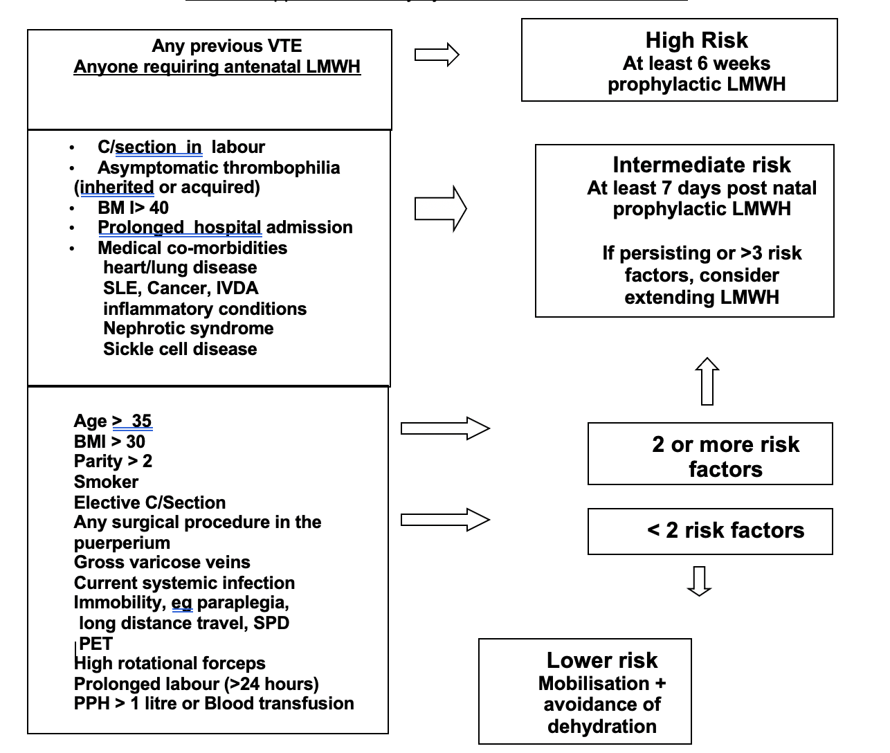
Gestational thrombocytopenia and indications for platelet counts in labour

Thrombocytopenia is usually defined as a platelet count under 150 × 109/L; this value represents the 2.5th percentile of the distribution in a healthy population of men and non-pregnant women.

The 2.5th percentile for the platelet count at the end of pregnancy (116 × 109/L) is significantly lower than the value accepted in the general population. This 2.5th percentile is similar for pregnant women of all nationalities or races; Europeans (North, South & Eastern), Asians, Africans, N.America & S.America.  Less than 1% of normal pregnancies have a platelet count of <100 x 109/l.

It is a commonly held belief (Bromage) that epidural / spinal block is safe with platelet count > 100 x 109/l. because as a (poor) predictor of surgical bleeding the bleeding time does not become prolonged until platelet count is < 100 x 109/l.

Gestational thrombocytopenia is common and thought to be due to hemodilution and or increased platelet turnover:

(Rolbin USA)

0.4% healthy pregnant women         - platelet count < 100 x 109/l

6.4%                                                   -      ..          ..     < 150 x 109/l 


(Burrows USA)

8.4% healthy women in labour         - platelet count 100 – 150 x 109/l


In Europeans, a similar prevalence of gestational thrombocytopenia exists (5.9% Finland, 10.9% Switzerland).

Population studies in healthy, normotensive pregnant women have repeatedly showed platelet count pre-pregnancy and in first trimester is normal, it falls through pregnancy with a nadir at 32-36 weeks gestation and then normalises at day 3-5 post-partum. The PT & APTT and bleeding time are normal. 

Hence in normal pregnancy (normotension and without proteinuria) in normal, asymptomatic women i.e. no history of easy bruising, recurrent epistaxis or prolonged bleeding after simple cuts or operations, not taking drugs or herbs nor suffering from a medical condition associated with a coagulation or haemostatic deficit a platelet count is not routinely indicated prior to performing epidural or spinal anaesthesia. There is a concern that ITP may be missed. This is unlikely, however, as all pregnant women attending for antenatal care get a full blood count performed at the booking clinic in the first trimester and again in the second trimester. Possible ITP would be noted from these tests. 

In the @ 0.4% normal pregnancies where the platelet count is < 100 x 109/l it would be reasonable to perform a platelet count, PT & APPT prior to performing an epidural or spinal. Finally, for those women who have not had routine antenatal blood tests performed, a platelet count prior to regional analgesia is indicated.

Gestational thrombocytopenia is not considered below a threshold of 75 × 109/L, where the diagnosis of HELLP, ITP, sepsis (chorio-amnionitis), auto-immune disorders, thrombotic thrombocytopenic purpura (TTP) should be considered.

In the group of women with thrombocytopenia between 75 and 115 × 109/L, @ 75% have HELLP or pre-eclampsia. In this group, there will be a clinical history and supportive BP recordings and urine proteinuria to confirm the diagnosis. Also consider HIV and Hepatitis C in women with similar level of thrombocytopenia but without proteinuria and hypertension.

In pregnant women with a platelet count less than 75 × 109/L, further investigations are essential. (PT, APTT, LFT’s, U&E’s)


Indications for platelet count & coagulation screen in labour
  1. History of congenital or acquired bleeding disorder
  2. Conditions assoc. with haemostatic dysfunction i.e. renal, hepatic, auto-immune disease
  3. Acute peri-partum conditions:
  • PIH or pre-eclampsia (BP 140/90 mmHg and proteinuria 2+)
  • HELLP
  • Placental abruption
  • Amniotic fluid embolism
  • Massive haemorrhage
  • Sepsis
  • Intrauterine fetal demise (risk increases from time of fetal demise)
  1. Monitor anti-coagulant therapy
  2. Monitor coagulation therapy e.g. Platelet transfusion in ITP, Clotting factor replacement in haemophilia, Von Willebrand’s disease
  3. No antenatal care.

In many of the above it is clear that coagulation tests are indicated and neuraxial blockade contraindicated anyway.

The incidence of spinal or epidural haematoma is rare @ 1/150,000 and is invariably associated with a coagulopathy i.e. PT, APTT are prolonged and other indices of coagulation are deranged (e.g. increased FDP, fibrinogen, D-dimer). There is also a cause for the coagulopathy. Concomitant use of drugs affecting coagulation (warfarin, heparin or LMWH) and old age are other associated factors.

Idiopathic thrombocytopenic purpura

Commonly presents in young women - 1/1,700 prevalence in pregnancy. Differentiating these from normal pregnancy with low platelet count is the problem as @ 66% ITP are mild and asymptomatic during pregnancy. Pointers are a low platelet count pre-pregnancy and 1st trimester of pregnancy (75-100 x 109/l). Symptoms when present range from simple bruising, purpura, through epistaxis to GI bleeding. Possible family history of ITP or other auto-immune disease esp. SLE may be found. Individual platelet function maybe normal or increased. Platelet count tends to fall further during pregnancy. Case reports of inadvertent or deliberate but uncomplicated epidural or spinal injections in patients with platelet counts < 20,000 x 109/l. exist.


Dr S Rowbottom

May 2005

(updated Dr P Purvis 2022)

References

  1. Rolbin SH. et al. Epidural anaesthesia in pregnant patients with low platelet counts. Obstet Gynecol. 1988;71:918-920
  2. Burrows RF. N Eng J Med 1988:319(3);142-5. (July 21)
  3. Sainios S. Acta Obstet Gynecol Scand. 2000;79:744-9
  4. Boehlen F. Obstet Gynecol. 2000:95;29-33
  5. Bromage PR. Neurological complications of regional anaesthesia for obstetrics. In Shnider SM. Anaesthesia for Obstetrics.
  6. Burrows RF, Kelton JG. Fetal thrombocytopenia and its relation to maternal thrombocytopenia. N Engl J Med 1993;329:1463–6.
  7. Boehlen F, Hohlfeld P, Extermann P, de Moerloose P. Maternal antiplatelet antibodies in predicting the risk of neonatal thrombocytopenia. Obstet Gynecol 1999;93:169–73.

Inadvertent dural puncture

Once the diagnosis has been made, it is important that successful analgesia is achieved. There are two alternative strategies:

1) Re-site the epidural in an adjacent (cranial) space

This can be done by yourself if you are sufficiently confident, or by a more senior anaesthetist. Don’t be surprised or worried if your confidence falters after doing an inadvertent dural tap.

Be cautious with the first dose of local anaesthetic through the catheter as the tip may lie near the puncture site. All doses should be fractionated and given over 3 to 5 minutes.

2) Continuous subarachnoid block

This must be discussed with a senior colleague.

Catheter (preferably end hole) is passed into the subarachnoid space. 2 cm only should be left in the space. The catheter must be clearly marked as being subarachnoid’ or ‘spinal’.

1 ml initially, then 0.5 ml increments, of the standard solution (0.1% levobupivacaine with 2 mcg/ml fentanyl) can be used to establish analgesia and for subsequent top-ups. Doses should be given with the patient in the lateral or supine wedged position. All doses must be given by an anaesthetist.

It is important to appreciate that levobupivacaine is slightly hypobaric thus sudden movement risks unpredictable displacement in the CSF, and sitting up may result in an unexpectedly high block.

For LSCS, manual removal and rotational forceps delivery, with the patient in the lateral or wedged supine position, give 0.5 ml increments of 0.5% heavy bupivacaine until adequate anaesthesia is achieved. A maximum dose of 4 mls should be given as caudal pooling of solutions containing dextrose has been associated with development of cauda equina syndrome.

For lift out forceps, positioning as above, give 1 to 2 mls of 0.5% heavy bupivacaine.

General points

Once analgesia is established a full explanation should be given to the patient, her partner and the midwife looking after her. The obstetric team should be informed.

Elective forceps delivery is no longer considered essential but having allowed adequate descent of the presenting part, excessive pushing/straining should be avoided. Obviously, definition of excessive is an obstetric clinical judgement.

After delivery, the mother should be allowed to mobilise freely. Restricting her to bed rest will not prevent PDPH.

Events must be clearly documented in the notes and the patient visited daily until she is discharged. She should be given labour ward contact details, a letter for her GP and a RCOA ‘Headache post epidural’ information sheet (available in the labour ward anaesthetic room). The on-call consultant should be informed.

Post dural puncture headache (PDPH)

70 - 90% of women who have an inadvertent dural puncture with a 16G Tuohy needle will develop a PDPH. The incidence is < 1% with specially designed spinal needles. Parturients are more susceptible to PDPH than the general population. Symptoms usually start within 48 hours of the dural puncture and last approximately 10 days, but may last weeks, months or rarely years.

The pathophysiology is not entirely clear, but PDPH is thought to occur when leak of CSF leads to contraction of the subarachnoid space and compensatory expansion of pain sensitive intracerebral veins, cranial nerve roots and/or meninges.

Typically the mother has a severe, disabling fronto-occipital headache, which is relieved by lying down and worsened by standing or sitting up. The headache may radiate to the neck/shoulders and be associated with other symptoms such as nausea, visual disturbances and even photophobia. If the patient sits up and upper abdominal pressure is applied the headache will often temporarily improve, supporting the diagnosis.

The symptoms are distressing and usually prevent the mother from being able to care properly for her newborn child. Immobility increases the risk of thromboembolic disease and infection, and discharge from hospital is usually delayed. More severe complications such as cranial nerve palsies, subdural or intracranial haemorrhage can occur, but are thankfully very rare.

A full history and neurological examination should be performed. The following alternative differential diagnoses should be considered and excluded:

  • simple headache or migraine
  • meningitis (septic or aseptic)
  • preeclampsia
  • electrolyte disturbance or hypoglycaemia
  • raised intracranial pressure
  • central venous sinus thrombosis.

Conservative management:

  • simple analgesics (paracetamol & nonsteroidal anti-inflammatories)
  • avoid dehydration
  • avoid constipation (may need lactulose).
  • caffeine – 300mg in 24h

Dehydration can exacerbate PDPH so should be avoided. Overhydration does not prevent PDPH.

Other measures such as sumatriptan and sphenopalatine ganglion block have been used but there is no strong evidence for their efficacy. As such, their use in our unit is not routine.

If the PDPH does not resolve within 24 hours with conservative management an epidural blood patch should be discussed and offered.

Epidural blood patch (EBP)

This is the definitive treatment for PDPH occurring following inadvertent or deliberate dural puncture. The reason or way it works is unclear, although it is thought to relate to effects on cerebrospinal haemodynamics.

Prophylactic (ie postpartum, just before removal of epidural catheter) blood patching has been used but there is no evidence to support its use.

EBP should be performed after full explanation to the mother and full discussion with the consultant on for labour ward. A consent form should be signed by the clinician and the patient.

Ensure the patient is apyrexial and that other causes for severe headache have been excluded.

Ensure also that the patient has not received LMWH within 12 hours.

Contraindications are as for other central neuraxial procedures (see Section 1.2).

Side effects include failure, inadvertent dural puncture, back pain, transient nerve root pain and pyrexia.

EBP is effective in > 70%; headache may recur in up to 50% of mothers necessitating a further blood patch. When successful, relief is usually immediate.

Procedure

This requires two operators using aseptic techniques.

One operator sites the epidural needle in the epidural space at, or just below the level of the puncture.

The second takes 20 mls of autologous blood from the patient under sterile conditions.

The blood is injected slowly via the Tuohy needle into the epidural space, stopping if the mother complains of pain in her back or legs (a sense of ‘fullness’ is often expressed).

2 mls of saline should be used to flush the epidural needle as it is withdrawn so that a plug of blood is not left.

The mother should lie flat for 2 hours then be encouraged to mobilise normally.

Hypotension

It is uncommon to get significant hypotension from a correctly managed epidural block for labour. The possibility of aortocaval compression, inadvertent subarachnoid or subdural block should be considered.

If however, the blood pressure falls (< 100 mmHg or > 30 % fall from pre-block mean arterial pressure) move the mother into a full lateral position. Infusion of a small volume (100 mls) of crystalloid and increments of 3mg ephedrine or 20mcg phenylephrine should be given intravenously to restore normotension. Pre-filled syringes of ephedrine are kept on the epidural trolley.

Beware of giving large volumes of crystalloid to women in labour, particularly if labour is being augmented with syntocinon; syntocinon has an antidiuretic effect and the combined result may be water intoxication, hyponatraemia and convulsions. A careful record of fluid balance should be kept through labour.

 Pruritus

More common with neuraxial than systemic opioids and in pregnant versus non-pregnant women (probably effect of oestrogen on opioid receptors). Severe in < 1%. Mechanism not entirely clear. Incidence is highest with morphine and lowest with the more lipophilic drugs eg fentanyl.

If the patient is distressed and treatment is needed, the following can be tried:

  • simple antihistamines
  • naloxone bolus (40 mcg iv increments)
  • naloxone infusion (0.4 – 0.6 mg/h)
  • other treatments such as propofol, ondansetron, nalbuphine & promethazine have not been formally evaluated.

High block & inadvertent (total) spinal

A high block can be defined as spread of a block three or more segments higher than anticipated.

A total spinal can be defined as a block that results in loss of consciousness and severe cardiorespiratory compromise.

May be due to inadvertent subarachnoid (> 1: 5,000 - 50,000) or subdural (up to 1: 100 obstetric epidurals) administration of local anaesthetic or just unpredictable spread.

Careful epidural administration of fractionated doses of local anaesthetic should help identify an inadvertent spinal and avoid precipitous changes in block height.

Subdural blocks are characterised by slow onset (approximately 20 minutes) of a high but patchy sensory block, often with poor analgesia and usually with little motor block or hypotension. The epidural catheter should be removed and the epidural resited.

For high block (> T4) with cardiorespiratory compromise but no loss of consciousness:                            

  • call for help if you need it
  • place in full left lateral position
  • give oxygen via a facemask (> 10 l/min)
  • give 3 -6 mg boluses of ephedrine or 20-40mcg of phenylephrine
  • atropine 600mcg or glycopyrollate 200mcg likely to be required if cardio-accelerator fibres involved (T1-T4)
  • give 500 ml bolus of fluid
  • reassure mother and her partner
  • observe mother and foetus closely.

For total spinal as described above:

  • call for senior anaesthetic and obstetric assistance (2222, state “Obstetric Emergency” and location)
  • place in full left lateral position
  • give 100% oxygen with bag & mask if apnoeic
  • give 500 – 1000 ml IV fluid rapidly
  • give 15 mg ephedrine IV and repeat as necessary
  • Intubate if unconscious & apnoeic
  • put full monitoring on
  • consider using other vasopressors (eg phenylephrine in 50 - 100 mcg increments or even adrenaline in 5 – 10 mcg increments if hypotension resistant)
  • consider emptying stomach with an orogastric tube
  • monitor fetus and consider delivery once mother stabilised.

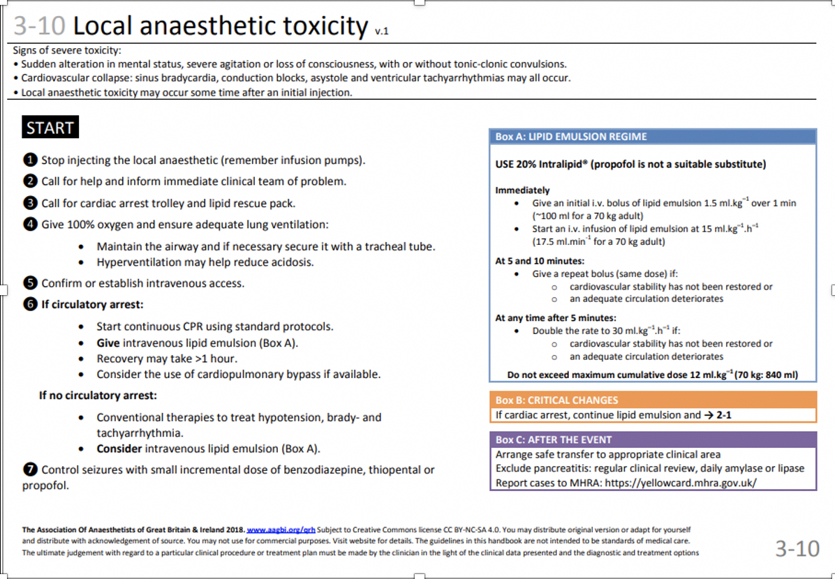
Local anaesthetic (LA) toxicity

This may manifest itself with predominant central nervous system (CNS) or cardiovascular (CVS) effects. Of the agents most commonly used on labour ward, the former is more commonly seen with lidocaine, and the latter with bupivacaine.

CNS irritability resulting in convulsions may progress to CNS depression; CVS collapse can lead to cardiac arrest.

Symptoms and signs may include: tinnitus, light-headedness, visual disturbances, circumoral numbness, drowsiness, convulsions, apnoea, hypotension, ECG changes (­P-R interval, A-V dissociation, ­QRS), sinus bradycardia and asystole.

Newer agents (ropivacaine & levobupivacaine) seem to have a wider safety margin compared with bupivacaine, which behaves in a particularly cardiotoxic way in pregnant women.

LA toxicity in this setting usually occurs where excessive doses of LA have been used (epidural administration), or where inadvertent intravenous injection has occurred.

The following dose limits should not be exceeded:

Bupivacaine/Levobupivacaine              2 mg/kg

Lidocaine                                                 3 mg/kg without adrenaline

                                                                 7 mg/kg with adrenaline


All doses should be fractionated and given only after careful aspiration.

If suspected LA toxicity occurs:

  • stop giving LA
  • call for help (anaesthetic & obstetric)
  • ABC pattern of resuscitation (NB remember tilt/wedge to avoid aortocaval compression)
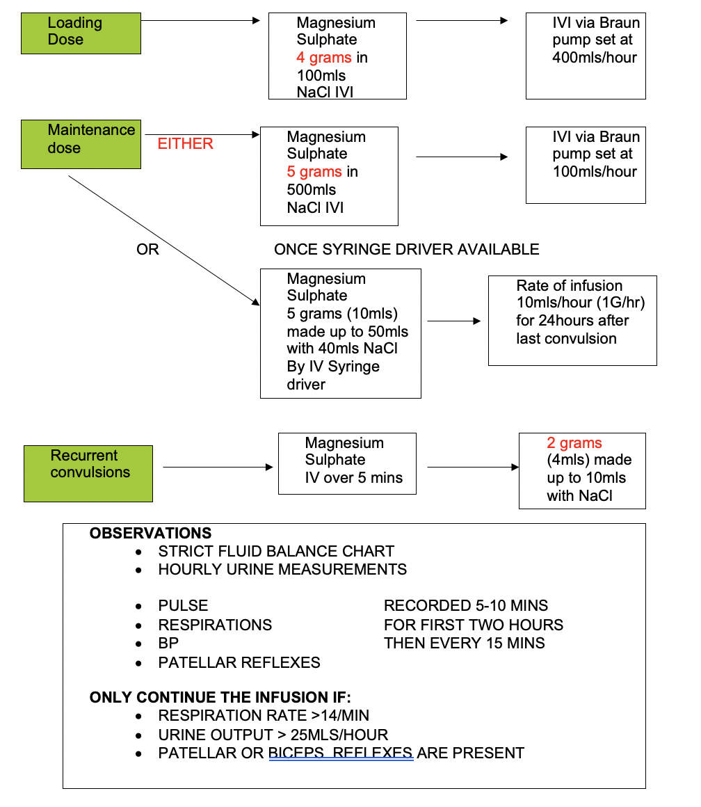
  • treat convulsions with diazepam (5 -10 mg IV) or thiopentone (50 mg increments)
  • consider giving intralipid. This is stored in the labour ward theatre cupboard next to the local anaesthetic agents. It is also kept in the arrest trolleys, both in labour suite and main theatre recovery. See page 57 for recommended treatment regime.
  • The LA toxicity guideline in the Association of Anaesthetists Quick Reference Handbook can also be used.


Problems on post-natal follow-up

The anaesthetic team on labour ward is responsible for post-natal follow-up of all women who have had a peripartum anaesthetic procedure. Usually the consultant does the visits, but trainees are also expected to do them, particularly at the weekend, unless exceptionally busy.

The post-natal visit serves several purposes:

  • prompt detection of complications of anaesthetic procedures
  • opportunity to discuss experience (particularly if unpleasant)
  • collection of audit data.

In addition to questions regarding efficacy of and satisfaction with, analgesia /anaesthesia, information regarding the following should be sought:

  • neurological symptoms & signs (including bladder/bowel sensation & function)
  • pain control
  • nausea/vomiting
  • pruritis
  • backache

If in doubt, take a full history, perform an examination and discuss the patient with the on-call anaesthetic consultant covering the unit.

These mothers must contact one of the obstetric anaesthetic consultants (via the community midwife) if the headache recurs or there are any other adverse neurological symptoms.


Anaesthesia for Caesarean Section

Degree of urgency & choice of technique

Good communication between patient, anaesthetist, obstetrician, midwife and theatre staff is essential.

Broadly speaking, four situations exist:

Category 1:  Immediate threat to life of woman or foetus (eg major antepartum haemorrhage, cord prolapse, placental abruption). Delivery should be as soon as possible, and in most situations within 30 minutes of making the decision.


Category 2: Delivery should be as soon as possible and may be communicated as an urgent Cat 2 in some circumstances. 

If a working epidural is in place, a top-up can be given (see 3.5). A spinal can also be used, but the anaesthetist must not delay surgery for repeated attempts if difficult. A regional technique is nearly always the technique of choice, but if the above fail within the given time frame, or if the obstetric staff insist that immediate delivery is needed, then a general anaesthetic is appropriate.


Category 3:     Needing early delivery but no maternal or fetal compromise.


Category 4:     At a time to suit the patient and maternity team (Elective C/S).


The anaesthetic of choice is a spinal, either as a single shot or as part of a combined spinal epidural technique. Spinals are quicker onset, denser blocks which give better operating conditions than epidurals. Only a small dose of local anaesthetic is required and the risk of headache is low (< 0.5%) when a pencil point needle is used. If there is a contraindication to regional techniques a general anaesthetic will be needed.

General points

Remember, for spinals, epidurals or CSEs:

All patients should be assessed as for a general anaesthetic and should receive antacid therapy (see p40)

A block from T4 to S5, (testing modality = temperature), is needed for CS. We also feel that using touch as the sensory modality; the block should extend to T6.

The block must be tested and the following details recorded:

  • time of block
  • time block tested
  • height of block & sensory modality used to test it (ideally document motor block, block to cold and block to light touch)
  • times of incision & delivery
  • any comments on block quality or any treatment offered per-operatively (bottom-left corner on the front page of the anaesthetic chart).

Explain as fully as possible what you are going to do, and what the patient can expect to see, feel and hear.

Reassure and warn patient that if an adequate block cannot be performed, or an unusual surgical situation is encountered, then a general anaesthetic may be needed.

Give syntocinon 5 IU as a slow intravenous injection once the anterior shoulder is delivered – you do not need to wait until the cord is clamped. The exception being twins/triplets where syntocinon is given after delivery of the final twin’s anterior shoulder.

The cord will then either be clamped immediately or will be followed by a 1-minute delay, depending on the clinical situation. There should be a discussion with the Obstetrician whether they would like additional uterotonics given. It is a dynamic process and therefore ongoing dialogue should continue between the Anaesthetic and Obstetric team throughout the Caesarean section.

Good documentation is essential!

(See “Medical and Legal Aspects of Regional Block for Caesarean Section” JH McClure at the end of this handbook)


Blood Pressure Maintenance with Phenylephrine Infusions

(NB This guideline does not cover the use of phenylephrine infusions in any other cases but uncomplicated deliveries)

Introduction

Phenylephrine, a directly acting alpha agonist used for the treatment of hypotension, has been shown to be a useful vasopressor in obstetric anaesthesia and does not cause a decrease in fetal cord pH compared to other agents. As it has a relatively short half-life, it lends itself to administration by continuous iv infusion when treating hypotension induced by regional or general anaesthesia for operative / assisted delivery.

Indications

  • Hypotension following regional anaesthesia for assisted/ operative delivery
  • Hypotension following general anaesthesia for operative/ assisted delivery

Contraindications

  • Known sensitivity to phenylephrine
  • Hypertension (relative)
  • Severe pre-eclampsia/ eclampsia (relative)

Preparation

  • Inject 10mg of phenylephrine into a 500ml bag of 0.9% NaCl solution- final concentration 20mcg ml-1
  • Draw up 50-60ml into a syringe fitting Alaris pump
  • Load the syringe into the syringe driver and connect to the patient’s iv access via a y connector and extension
  • Use the ‘purge’ or ‘bolus’ function on the driver to flush the extension prior to connecting
  • Always administer together with a running iv infusion

Regime

      For operative or assisted delivery under spinal anaesthesia

  • start infusion at a rate of 60 - 90 ml hr-1 immediately after injecting spinal drugs
  • vary infusion rate according to clinical response up or down by 30-10 ml hr-1

For operative or assisted delivery under top up epidural anaesthesia or general anaesthesia

  • start infusion at a rate of 30 - 90 ml hr-1if clinically indicated
  • if phenylephrine is required consider giving a bolus of 40mcg (2ml) before infusing
  • vary infusion rate according to clinical response up or down by 30-10 ml hr-1

Use pump to administer 20 or 40 mcg boluses as indicated

The infusion can usually start to be weaned following delivery (maternal auto-transfusion and relief of any aortocaval compression), most infusions will be discontinued after 45 minutes. If still requiring high doses of vasopressor, think – is there another cause of hypotension (i.e. haemorrhage)?

Spinal anaesthesia or subarachnoid block

PREOPERATIVE DISCUSSION

The following should be discussed in full with the patient:                     

  • how the procedure is performed
  • rapidity of block onset
  • possibility of hypotension with feelings of light-headedness & its treatment
  • tugging, pulling & pushing sensations
  • possibility of peroperative pain & its management
  • possible complications of spinal anaesthesia including failure, nerve damage and headache.
  • failure of anaesthesia is surprisingly common (1:50 spinal, 1:20 epidural top-up), moreso than many other complications for which we consent. It leads to significant morbidity (distress, conversion to GA), and there are specific stickers you should append to the anaesthetic chart to confirm this has been discussed (see below).

PREPARATION OF PATIENT

Attach monitoring (electrocardiograph (ECG), non-invasive blood pressure (NIBP) & pulse oximeter) and record baseline vital signs.

Insert at least a 16g intravenous (IV) cannula preferably in the non-dominant forearm/hand, using local anaesthesia.

Flush cannula and attach a 1000 ml bag of PlasmaLyte 148 solution.

USE AN ASEPTIC TECHNIQUE

Ensure back sprayed with 0.5% Chlorhexidine spray before spinal pack opened. Scrub hands and arms; wear protective/aseptic clothing (hat, mask, gown & gloves)

Prepare equipment aseptically (swabs, 25 or 27 G spinal needle & introducer, local anaesthetic solution to initiate block).

CHOICE OF ANAESTHETIC AGENT

Hyperbaric bupivacaine is the local anaesthetic used; doses of 10.6 –13.5 mg (2.3-2.7 ml of 0.5% solution) have been shown to be sufficient. 2.5 mls (12.5mg) heavy 0.5% bupivacaine is a safe dose. Opioid may be added to this to improve the quality of the block. The routine in St. John’s obstetric department is preservative free diamorphine 0.3 mg (available in sterile, double-wrapped ampoules). This gives excellent extended analgesia postoperatively, but carries a very small risk of sedation and late onset respiratory depression. The Obstetric SEWS chart has a section indicating that the patient has had intrathecal opioids; this section MUST be completed when opioids are used.


In the event of diamorphine shortages, fentanyl 12.5 mcg (0.25 mL preservative-free) is a relatively safe choice


The procedure (mid-line approach)

We have a SOP for carrying out spinal anaesthesia for elective Caesarean Section, which is appended at page 80 of this handbook. This stresses the need to confirm (with the ODP assisting) verbally and visually both the intended local anaesthetic and opioid drugs, and the preparation of these drugs as the injectate for the spinal.

  • Ensure patent large-bore IV access (18G at a minimum) before starting. A cannula sited earlier in labour may have become displaced or blocked.
  • Place patient in preferred position (sitting or right lateral). Inform patient of all further manoeuvres before/as they happen.
  • Ensure the Chlorhexidine has had time to dry. Place sterile drape to cover back & hips.
  • Palpate the hips to establish position of interspaces (Tuffier’s line, drawn between the iliac crests, usually, but not always, crosses the L3/4 interspace. It lies slightly higher in the pregnant state, because of pelvic rotation). Using this as a guide select a suitable space for needle insertion - preferably L3/4 or L4/5.
  • Use lidocaine 1% or 2% to anaesthetise area around insertion site. Avoid using a green needle as these have been known to cause accidental dural punctures during skin infiltration.
  • Insert spinal needle slowly & carefully - If any paresthesia or pain occur, note this, stop, remove needle and reinsert.             
  • A ‘give’ is usually felt as the needle traverses the dura.
  • The stylet should then be withdrawn and free flow of clear cerebrospinal fluid (CSF) should be seen.
  • Attach the syringe containing the local anaesthetic, confirm free aspiration of clear CSF, and then slowly inject the dose.
  • Once the injection is completed, immediately lay the mother in the left lateral position using a wedge or table tilt.
  • Confirm that the drip is running and put the NIBP to record every 3 minutes.
  • Note the time of the block, and after 5 minutes, test both sides using ethyl chloride and/or cotton wool.
  • Allow the CS to start only when you are happy there is a dense bilateral block from T4 to S5.
  • Document the details on the anaesthetic chart, the audit record and the MEWS chart.


HYPOTENSION DURING CAESAREAN SECTION

Hypotension due to spinal anaesthesia is common, and can cause nausea as well as placental hypoperfusion.  Make sure that the patient is adequately tilted, speed up IV fluids, and give a vasopressor.  Increments of ephedrine (3-6mg) or phenylephrine (20-40mcg) can be given. If phenylephrine is given first, beware of bradycardia-glycopyrrolate (200mcg) may be needed.

EPIDURAL ANAESTHESIA FOR LSCS

Not the first choice for CS, epidural anaesthesia is usually used where there is an epidural in situ for labour.

See page 9 for guide to insertion of an epidural catheter for obstetric analgesia/anaesthesia

Pain/discomfort and ‘missed segments’ are more common with epidurals compared with spinals and this should be discussed with the patient. It makes the careful assessment of the block prior to commencement of surgery, all the more important.

Hypotension is usually slower in onset and less severe than with spinal anaesthesia, but a free running intravenous cannula (16 G) is still essential and treatment with ephedrine or phenylephrine may be required.

Migration of the epidural catheter (subdural, subarachnoid or intravenous) is always a possibility thus epidural doses should be fractionated or given slowly, whilst monitoring the patient closely.            CHOICE OF DRUGS

20 ml lidocaine 2% + 1:200,000 adrenaline (slightly faster onset)

20 ml bupivacaine 0.5% or levobupivacaine 5mg/ml (slightly longer lasting block)

Diamorphine 3mg may be added to any of the above solutions to optimise block quality, and give up to 18 hours of postoperative analgesia. Alternatively, fentanyl 50 - 100 mcg will improve the quality of block and give 4-6 hours of postoperative analgesia. 

2ml 8.4% sodium bicarbonate has been used to ‘carbonate’ solutions and speed block onset. However, there has been concern regarding the risk of drug errors when mixing solutions and this is not routinely used.

Epidural local anaesthetic administration

A volume of 20 -25 ml of one of the above solutions is usually needed. Slow bolus injection has been shown to produce a more rapid reliable block compared with 5 ml increments every 5 minutes. Obviously, this goes against the gold standard for epidural top-up administration but if the urgency of the situation merits its use, the patient must be monitored closely to detect inadvertent intravenous or intrathecal injection. If time allows, incremental administration of the local anaesthetic dose is the preferred technique. 

Combined spinal & epidural anaesthesia (CSE)

We would not expect trainees at St John’s Hospital to undertake CSEs without prior discussion with a consultant.

May be needle through needle, or separate space technique

In theory this technique offers the reliability and speed of onset of a spinal with the flexibility and continuity of an epidural.

Although not routinely used in our unit, the CSE may be appropriate where complex/prolonged surgery is anticipated but it is still possible for the patient to be awake.

Two different techniques are commonly described

  • normal spinal dose with epidural for back up and/or postoperative analgesia
  • smaller intrathecal dose of local anaesthetic (giving a block to T8 -T10) with gradual extension using the epidural catheter. This offers better haemodynamic stability compared with the former.

Potential problems/complications with a CSE include:

  • there is a higher failure rate of spinal component (improves with experience)
  • the epidural catheter is untested at time of insertion and careful incremental top-ups must be used
  • there is possibly a slightly higher risk of meningitis, neuropraxia and neurological damage than with either technique alone, although evidence is not conclusive.

Management of spinal anaesthesia in the presence of inadequate epidural analgesia for urgent Caesarean section

Inevitably, and even in the best of hands there is a failure rate for epidural injections.

Unrecognised partial dislodgement of epidural catheters and persistent unilateral epidural blocks are possibly the main causes of an inadequate sensory block for a Caesarean section following an epidural top-up via a previously inserted epidural catheter. 

In the situation where following an epidural top-up of motor blocking local analgesics (e.g. 15 ml of 2% lidocaine in 1/200,000 adrenaline or plain 0.5% (levo-) bupivacaine) that there is an inadequate sensory block present for surgery to proceed; the options are to either give a general anaesthetic or administer a spinal anaesthetic.

This decision depends on the urgency of the situation and any obvious anticipated difficulties with airway management.

Be very careful about giving spinal anaesthesia in the presence of a partial sensory block and a recent (<30 mins.) epidural top-up. The same advice also applies to a recent epidural top-up of low concentration local analgesic solutions with added opiate for analgesia in labour.

This is a controversial area as there are many case studies reporting unpredictably high block and in some cases total spinal block with cardio-respiratory arrest when conventional doses of spinal anaesthetic are given. This is thought to be due to the volume compression effect on the CSF by the recent epidural top-up and that partially anaesthetised spinal nerve fibres require less local spinal analgesic solution and there is an excessive spread of the residual. Conversely, there are reports of uneventful spinal anaesthesia following the use of conventional doses of spinal analgesic in the same scenario!

One needs to distinguish between those cases where;

(a) Following the epidural top-up with an appropriate volume of local analgesic (of motor blocking concentration) there has been an increase in the density of the block but it will not ascend high enough for surgery i.e. the epidural catheter is at least partly in and delivering LA solution to the epidural space;

(b) There is no discernible increase in the density of the sensory or motor block following an appropriate top-up, i.e., the epidural catheter is no longer in the epidural space.

If (a) is the case, discuss with a senior or consultant anaesthetist, consider reducing the dose of spinal anaesthetic and proceeding as below. The decision to reduce the dose of spinal anaesthetic will be guided by the existing level (pre-spinal) of the sensory block.

  • perform spinal anaesthesia in the sitting position
  • reduce the dose of spinal anaesthetic solution by up to one third
  • omit intrathecal opiates if epidural / parenteral opiates already given
  • keep the patient sitting for @ 1minute before changing position for surgery
  • ensure vasoconstrictors are prepared and immediately available.

If (b) is the case, proceed with spinal anaesthesia using conventional doses as appropriate to weight & height.

Dr S Rowbottom

July 2005

(updated Dr P Purvis May 2022)Management of an inadequate regional block

Surgery should not commence until a dense motor and sensory block from T4 to S5 has been demonstrated.  But, what if:

You cannot perform regional anaesthesia

  • seek senior assistance as soon as difficult regional anaesthesia becomes apparent
  • general anaesthesia may be necessary – communicate with the woman, senior obstetrician, and midwife regarding urgency of the situation

The patient complains of pain intraoperatively

  • if epidural in place, ask the surgeon to wait and give a top-up of local anaesthetic +/- opioid (according to how much has already been given).
  • consider small incremental doses of intravenous opioid (if post delivery) but be wary of causing excessive sedation. Alfentanil has a rapid onset and may be useful.
  • consider trying N2O/O2
  • a general anaesthetic may be needed if the patient does not respond promptly to one of these measures, and should be offered if the patient is distressed.
  • any pain experienced and any action taken/treatment offered must be clearly recorded on the anaesthetic chart.
  • intravenous midazolam is not an appropriate treatment for an inadequate regional block.

Don’t struggle on if you are finding the spinal/epidural difficult to do: ask for help.

It is particularly important to ensure that patients who have had a difficult time for some reason are adequately followed up, with an opportunity to discuss events and any implications that there may be for future anaesthesia/pregnancy.

General anaesthesia

More than 80% of Caesarean sections (CS) are performed under regional block, in recognition of the potential morbidity and mortality associated with use of general anaesthesia in the obstetric population. The main concerns are

  • potentially difficult airway (1:300 failure to intubate)
  • increased risk of pulmonary aspiration in pregnant vs non-pregnant
  • risk of awareness (minimal with modern techniques & monitoring).

Indications for general anaesthesia are relatively few: technical failure of regional technique (this should hopefully be rare) contraindication to regional technique

  • patient refusal
  • sepsis (local or generalised)
  • haemodynamic instability
  • raised intra-cranial pressure
  • abnormal coagulation (pathological or iatrogenic)
  • surgical indication (eg anterior placenta praevia with previous CS)
  • obstetric indication (eg severe fetal distress)

Preoperative assessment

This need not be too lengthy but is essential even in the most pressing circumstances. It should include questions regarding relevant medical & obstetric history, previous anaesthesia, drug allergy, food intake and indication for, and urgency of CS.

The airway must be assessed (mouth opening, neck & jaw movements) as well as cardiorespiratory status in the presence of maternal compromise.

Premedication

Omeprazole 20mg PO (may already have been given in labour; see Kardex)

0.3 M sodium citrate 30 ml PO, immediately before preoxygenation

A dose of IV cefuroxime 1.5g is the routine antibiotic prior to skin incision.  If history of anaphylaxis to penicillin, consider giving clindamycin.

Induction

Machines and drugs must be checked daily. A skilled assistant is mandatory, to apply cricoid pressure and assist with intubation.

  • free running, large bore cannula (16 or 14G) in situ
  • induce in theatre, with the patient wedged/tilted in the left lateral position
  • full monitoring (ECG, pulse oximeter, NIBP, capnograph attached to filter)
  • preoxygenate (3 minutes or 5 vital capacity breaths)
  • rapid sequence induction with thiopentone (5mg/kg unless haemodynamic problems) and suxamethonium (1 - 1.5 mg/kg)
  • other drugs may be used in certain situations (eg alfentanil in preeclampsia).
  • You may prefer to use propofol and rocuronium for induction based on previous experience. This is entirely acceptable, but these drugs should be prepared and stored separately in the drug fridge to avoid confusion.
  • cricoid pressure is applied as consciousness is lost, and maintained until the airway is secured and tracheal intubation confirmed.

Maintenance

Usually 50% nitrous oxide + 50% oxygen pre- and 70% nitrous oxide + 30% oxygen post-delivery, plus volatile.

Non-depolarising muscle relaxant (usually atracurium) should be given once the suxamethonium has worn off. A peripheral nerve stimulator should be used. Remember to check nerve twitches after giving suxamethonium before giving non-depolarising agents; suxamethonium has a prolonged duration of action in pregnancy due to reduced plasma cholinesterase activity. This also allows for detection of rare but important suxamethonium apnoea.

Oxytocin 5 IU is usually given as a slow IV bolus after delivery of the anterior shoulder. If requested by the obstetricians, this may be followed by 40 IU in 500 ml 0.9% NaCl over 4 hours.

Adequate analgesia is essential; IV morphine titrated to response peroperatively and diclofenac 100 mg PR (unless contraindicated, consent gained preoperatively) at the end of the operation are usually used. Regional anaesthesia of the abdominal wall may also be helpful and many consultants at SJH are proficient; discuss with the consultant on for Labour Suite.

Recovery

Ensure residual neuromuscular blockade is adequately reversed (neostigmine 2.5 mg + glycopyrrolate 0.5 mg).

Wake the mother up in the left lateral position.

Extubate only once the mother is awake with intact protective airway reflexes.

Nurse in a properly staffed and equipped recovery area until ready to return to the observation area.

See analgesia guidelines (page 45).

Follow-up

An essential part of patient care anyway, but particularly important when patient has undergone emergency general anaesthesia.

A careful technique should avoid the risk of awareness, but reassurance and explanation of events may still be needed.


Management of failed intubation

A number of drills and flow charts exist- there is one on the wall in theatre (reproduced in the front of this handbook – Useful Resources). The only essential component is the maintenance of oxygenation. The other key factors are:

  • recognition of failure to intubate & calling for help
  • assessment of the airway & severity of difficulty
  • deciding whether or not it is appropriate to re-try.

The mother has to remain the anaesthetist’s priority; thus the mother should be allowed to wake unless:

  • surgery is life-saving for mother (eg major haemorrhage)
  • the decision is made by a senior/experienced anaesthetist.

Remember, patients die from failure to oxygenate not failure to intubate.


Perioperative Issues

Antacid prophylaxis

Acid aspiration is one of the factors associated with maternal morbidity and mortality and poses a threat when general anaesthesia is induced, or if conscious level is obtunded for any other reason (e.g. during a convulsion). Obstetric patients are at increased risk of aspiration of gastric contents when airway protection is lost, and of developing significant pneumonitis should aspiration occur. This increased risk is because of the following factors:

  1. Reduced Lower oesophageal sphincter tone
  2. Reduced Gastric emptying
  3. ­Production of acid (volume)
  4. Reduced pH of acid produced
  5. ­Intragastric pressure
  6. Physiological & physical difficulties with airway control & oxygenation

Prevention of acid aspiration is an important consideration in any woman more than 16 weeks pregnant or less than 24 hours post-partum. As with the non-pregnant population, symptoms of reflux indicate risk, irrespective of gestation.

Management strategy can be divided according to risk:

  • Normal/low risk labour: no antacid given routinely
  • High (obstetric or anaesthetic) risk labour: omeprazole 20mg PO 12hrly.

Elective CS: omeprazole 20mg PO on night before CS then omeprazole 20mg PO, approximately 2 hours pre-operatively.

Emergency CS: 20mg omeprazole PO at time of decision for theatre (may have received a dose recently if in labour).

 > 16 weeks pregnant or < 24 hours post-partum or symptoms of gastro-oesphageal reflux for other surgery: omeprazole 20mg PO, 2 hours pre-operatively.

In all of the above circumstances, if a general anaesthetic is to be given, 30mL of 0.3M sodium citrate should be given before induction (ie on arrival to theatre; it is kept on the airway trolley). 

Feeding in labour

Considerations

  1. During pregnancy:
  • lower oesophageal sphincter pressure is ¯ by smooth relaxation due to progesterone
  • áintra-abdominal pressure
  • árisk of pulmonary aspiration
  • 75 - 80% complain of heartburn during the 3rd trimester and a significant number will have a demonstrable hiatus hernia
  • there is some evidence that gastric volume is ­ & pH is ¯
  • gastric emptying is not delayed during pregnancy.
  1. During labour & delivery
  • gastric emptying is thought to be normal, unless opioids given
  • opioid analgesia by any route delays gastric emptying
  • gastric emptying & volume probably normal by 24 & 48 hours post-partum respectively
  • obesity, multiple pregnancy, polyhydramnios & lithotomy position all ­ risk of gastric reflux.

Isotonic drinks are rapidly emptied from the stomach, and absorbed from the gastrointestinal tract. Evidence supporting their use during labour is still not conclusive, but they remain a good option for fluid intake.

Antibiotic prophylaxis

Antibiotic prophylaxis for Caesarean section (CS)

  • Has been shown to reduce postoperative sepsis.
  • Usually given prior to skin incision.
  • Cefuroxime 1.5 g IV – prepared in 20mL water, slow bolus injection
  • Clindamycin 600mg if Penicillin allergic – prepare in 100mL bag of 0.9% saline and infuse over 30 mins.

Regional analgesia/anaesthesia in patients with sepsis

  • Risks cardiovascular collapse if significant systemic sepsis. There is also the risk of epidural and/or meningeal infection.
  • Will depend on the risk: benefit; discuss the case with a senior colleague if in doubt.
  • Ideally the decision regarding regional techniques in a pregnant patient with sepsis should be a whole-team discussion including the woman, anaesthetist, obstetrician and midwife. Risk should be individualised to the case wherever possible.

Thromboprophylaxis

Risk assessment for thromboembolism in parturients at time of delivery–

  • note this applies to delivery by Caesarean section and SVD

Dosing of LMWH for prophylaxis

Weight

Dalteparin

< 50

2,500 units - once daily

50-90

5,000 units – once daily

91 130

7,500 units – once daily / split

131-170

10,000 units – once daily / split

>170

75units / kg/ day once daily / split

Intermediate dose for 50-90

5,000 units twice daily

Duration Of treatment

Dalteparin should be commenced within 4-6 hours of delivery (allow 4 hours between removal of epidural catheter and first administration).

In most cases it should be continued for 7 days, with the last few days at home.

Patients with a past history of venous thromboembolism or underlying thrombotic problem should continue for a minimum of 6 weeks.

Specialist advice should be sought for those with a family history of thrombophilia.

Dalteparin is NOT contraindicated in breast-feeding.

Low molecular weight heparin & regional anaesthesia

Thromboprophylaxis- once daily dalteparin

  • Elective section – omit a.m. dose of dalteparin on day of section
  • Induction of labour – omit a.m. dose of dalteparin on day of induction
  • Labour epidural – if last dose of dalteparin > 12 hours ago, proceed with regional. If last dose < 12 hours ago, delay regional anaesthesia.

If patient requires emergency caesarean section within 12 hours of last dose of dalteparin, consult senior anaesthetist(s).  They may decide to use a spinal anaesthetic.

If a patient on LMWH presents in spontaneous labour within 12 hours of a prophylactic dose, they should not have an epidural. They may be offered an alternative form of     analgesia such as an opioid-based IV/PCA.

Thromboprophylaxis- on twice daily dalteparin

If the patient is on a B.D. dosage regime for thromboprophylaxis, the morning dose should be omitted on day of delivery.   They still present for regional within 12 hours of the last dose.

These patients constitute a high-risk group:

  • Consult with senior colleagues
  • Check clotting screen
  • A regional anaesthetic technique within 12 hours of dalteparin administration is relatively contra-indicated

Therapeutic Dosage

This dosage level is used for high-risk women.

This dosage regime has been associated with Vertebral Column Haematomas (VCHs), although usually in an older, possibly frailer patient. 

The administration of a regional anaesthetic technique within 24 hours of a therapeutic dose of LMWH is relatively contra-indicated.   Anti-Xa levels take longer to normalise.

Elective section – it is often possible to adjust the thromboprophylaxis and arrange to omit the dose of LMWH in the evening prior to the section date. Also omit the LMWH on the morning of operation, so that neuraxial blockade can be undertaken.  This would usually be planned at the combined haematology/anaesthetic high obstetric risk clinic in advance. Check coagulation screen, including anti Xa levels (need to discuss with haematology, sample goes to RIE).

Schedule for mid-morning to ensure 24 hours has elapsed since last dose.

Induction of labour – as above.  Ensure 24 hours has elapsed since last dose.

Emergency in labour – consult senior on duty.   Check coagulation screen (including anti Xa levels).  

If < 24 hours hours since last dose – relatively high risk.

If it is considered too high risk to omit 2 doses of dalteparin because of the risk of thromboembolism, general anaesthesia may be required for caesarean section.   Always check coagulation screen and discuss with senior anaesthetist.

Restarting dalteparin after Regional Anaesthesia

Wait at least 4 hours after regional anaesthetic before re-starting LMWH.  Peak plasma levels occur 3-5 hours after S.C. administration.   Remember these women are at high risk of thromboembolism so a greater delay is probably inadvisable; The risk of fatal PTE because of an omitted dose probably exceeds the risk of spinal haemorrhage with the combination of UH or LMWH.

Concomitant use of NSAIDs with dalteparin can increase the risk of bleeding – if in doubt as to whether to prescribe NSAIDs for post-procedural analgesia, discuss with the senior on duty.

Uterotonic therapy

Syntocinon is a synthetic oxytocin analogue, which acts directly on uterine syntocinon receptors to produce smooth muscle contraction. It is routinely used at CS to reduce the risk of postpartum haemorrhage (PPH). 5 IU should be given as a slow IV bolus following delivery. If requested by the obstetrician, the bolus dose may be repeated,  followed by an infusion over 4 hours of 40 IU syntocinon in 500 ml 0.9% NaCl solution (half life approximately 10 minutes).

Side-effects:

tachycardia & vasodilatation

antidiuretic effect

Thus care where:

fixed cardiac output states

prolonged therapy (do not dilute in water/dextrose)

Ergometrine has a direct contractile action on smooth muscle. It is most commonly used IM in combination with syntocinon during active (versus physiological) management of the third stage of labour. IV administration (up to 500 mcg, may be diluted)) may help stop PPH by increasing the force, frequency and duration of uterine contractions.

Side-effects:

vasoconstriction and hypertension (systemic & pulmonary)

increased CVP

nausea & vomiting

bronchospasm

increased ICP

Thus avoid where

hypertensive disease

significant cardiorespiratory disease

significant intracranial lesion

And care where:

patient is awake

Prostaglandins:

PGE1 (Gemeprost):    used to soften cervix prior to termination of pregnancy

PGE2 (Dinoprostone): most commonly used to induce labour (‘Prostin’)

PGF2a (Carboprost): used in PPH where standard oxytocics have been ineffective. 250 mcg IM is the usual starting dose and can be repeated per 15min, up to a maximum of 2g.

Side-effects:

nausea, vomiting & diarrhoea

hypertension

bronchospasm

flushing & pyrexia

Post-operative analgesia & antiemetic treatment

It is the anaesthetist’s responsibility to prescribe appropriate post operative analgesia after a Caesarean section as with all surgical procedures.

Appropriate analgesia usually includes:

  • Intrathecal or epidural diamorphine (remember to record its administration on the SEWS chart)
  • Regular oral paracetamol, 1g at tick times qds
  • PR diclofenac 100mg at end of surgery if no contraindications, and patient consents to this, then regular oral ibuprofen or oral diclofenac
  • MST, 20mg at 8 hours and 16 hours post-procedure
  • Oral morphine 10mg hourly as required, to a maximum of 60mg in 24 hours.

Note: Oxycodone is not a first line opiate, and should only be prescribed in specific circumstances. Slow release oxycodone (oxycontin) is not licensed for the first 24 hours post-operatively, and ward 11 does not stock it. If an oral opioid is required, then oral morphine solution should be prescribed.

Tramadol, oxycodone and codeine may all cause drowsiness in the breastfed infant.

Do not give NSAIDs where: severe pre-eclampsia (effect on platelet function), coagulopathy.  Care where:    history of asthma, (do not give if clear history of bronchospasm with NSAIDs or if have never taken any type of NSAID before), hypovolaemia or compromised renal function.

Analgesia after a GA Caesarean Section

After delivery of the fetus, an opiate should be given. These patients often need 15-20mg of IV morphine in theatre, and may need further IV morphine in recovery. If fentanyl is used, an adequate dose must be given.

If a GA is required for urgency but there is an epidural in-situ, consider giving epidural diamorphine. Post operatively, the above analgesic regime may be followed.

If an experienced operator is available then consider use of abdominal wall blocks (IINB/TAP/LQL).

Other as-required medicines

Antiemetics: Ondansetron 4mg IV 8 hrly is usually the first line anti-emetic in this hospital, followed by cyclizine 4 mg IV 8 hrly. If this fails, treatment with any drugs causing nausea eg opioids should be reviewed. Dexamethasone 4mg IV may be useful in patients who have persisitent problems despite ondansetron and cyclizine, although there is less experience with its use in obstetric practise.

All commonly used antiemetics carry a manufacturers ‘use with caution’ or ‘use only if essential’ in breastfeeding. Clearly it is a clinical decision based on risk versus benefit.

Laxatives: Should be prescribed to all women having Caesarean section or repair of perineal tears in theatre. Lactulose 10mL PO BD.

Antihistamines: for relief of opioid-related pruritus. 4mg chlorphenamine 8 hourly PO as required.

Peppermint water 10mL 6 hourly as required may also aid the relief of postoperative bowel spasm.

References

1.  Royal College of Obstetricians and gynaecology - Green-top Guideline, No.37a, November 2009.

Anaesthesia for other Obstetric Procedures

Pregnant patients presenting for other surgery

Pregnant women may present for surgery either related or coincidental to their pregnancy. Most are young and fit but the following must be borne in mind:

  • Diagnosis of abdominal conditions may be difficult, as may any abdominal surgical procedure, because of the gravid uterus
  • Obstetric conditions such as placental abruption and HELLP should be considered in the differential diagnoses where appropriate.
  • The risks of aortocaval compression, difficult airway management and aspiration of gastric contents must be remembered. The degree of risk will depend on the gestation, presenting problem and any coexisting medical condition.
  • The fetus is at risk from: effects of the primary illness
    • effects of any drugs used
    • effects on uteroplacental blood flow
    • effects on fetal oxygen delivery
    • premature labour provoked by any of the above

In general, surgery is avoided during pregnancy, particularly during the first trimester. Where it is unavoidable consider the following:

  • Careful pre-operative assessment and treatment with antacid therapy (after 16 weeks gestation)
  • Avoid the supine position, and use left lateral tilt particularly after 20 weeks gestation, to avoid aortocaval compression
  • Regional versus general anaesthesia should be weighed up for each individual case (NB volatile agents relax the uterus).
  • Most drugs are not licensed for use in pregnancy, use those ‘known to be safe’ and avoid newer drugs whose effects are unknown.
  • Avoid N2O (theoretical not proven risk)
  • Avoid drugs that might increase uterine tone (eg b blockers) and vasoconstrictors.
  • High maternal pO2 does not cause uteroplacental vasoconstriction; pCO2 should be kept in the normal pregnant range.
  • The fetus should be monitored pre- and postoperatively and if appropriate/possible, intraoperatively.

Manual removal of retained placenta (MROP)

This is required when the placenta has failed to deliver despite conservative measures (IM/IV oxytocics, turning into left lateral position, putting baby to the breast, emptying bladder). If surgical removal is required, regional anaesthesia is preferable either by top-up of an existing epidural or use of a spinal anaesthetic technique.

Anaesthesia should be adequate to allow manipulation of the uterus, thus a block in the region of T8 - T10 to S5 must be established.

There is a significant risk of major haemorrhage where the placenta/other products are retained. Adequate intra-venous access and resuscitation are an essential part of management. In the presence of ongoing haemodynamic instability, general anaesthesia (rapid sequence induction) is appropriate.

Have atropine/glycopyrollate prepared as, rarely, uterine inversion can complicate removal of retained placenta. Unless rapidly replaced the mother is likely to become hypotensive and bradycardic. In this circumstance, general anaesthesia may well be required. If the placenta/other products get trapped in the cervix a similar clinical picture may result from excessive vagal tone.

Repair of perineal tear

First- and second-degree perineal tears are usually sutured by the midwife under local infiltration/pudendal block. Repair of obstetric anal sphincter injury (OASI, i.e. third- and fourth-degree tears) requires obstetric input and transfer to theatre.

If an epidural is in situ it can be topped-up, aiming for a dense perineal block.

Otherwise, a spinal anaesthetic is the technique of choice. Keep the woman sitting upright for 30 seconds following injection to obtain a more dense “saddle block”.  A dose similar to that used for Caesarean section (2.5mL 0.5% heavy bupivacaine with diamorphine) is appropriate, and unlikely to result in a high block as there is now no gravid uterus to cranially displace CSF.

These cases are occasionally complicated by major obstetric haemorrhage due to vascular injury, and can require general anaesthesia. Obtaining an up-to-date estimate of blood loss and haemodynamic assessment during transfer to theatre is useful.

Trial of forceps

This term is used when it is anticipated that forceps delivery may be difficult, thus all must prepared for rapid conversion to Caesarean section. Thus it is usually carried out in theatre with regional anaesthesia suitable for either eventuality.

If there is a working epidural

Lidocaine 2% + 1:200,000 adrenaline can be used to rapidly provide a block suitable for Caesarean section, should that be required. 20-25ml is likely to be needed. 25 – 50 mcg of fentanyl can be added to optimise the quality of anaesthesia.

If there is no working epidural

Use a spinal anaesthetic technique where possible, using a dose suitable to provide anaesthesia for Caesarean section.

General anaesthesia should rarely be needed.

Instrumental delivery “in the room”

This may involve use of forceps (low, mid-cavity or high/rotational) or suction/Ventouse device. Indications for this type of delivery include:    

  • Prolonged second stage (>2 hrs for prim, >1hr for multip, add 1 hr if epidural in situ)
  • Maternal exhaustion
  • Failure of presenting part to descend
  • Contraindication to Valsalva manoeuvre
  • Fetal distress
  • Fetal immaturity

It is important to ensure a dense pelvic/perineal block. If there is a working epidural, use bupivacaine 0.5%, levobupivacaine 5mg/ml or lidocaine 2% + 1:200,000 adrenaline, depending on the urgency of the situation. A volume of 10 mls is usually sufficient to provide good pelvic anaesthesia, especially if the mother is kept in the sitting position. However, if there is ANY DOUBT that instrumental will be successful, top up as for Caesaerean section (see ‘trial of forceps’ above).

Shirodkar suture / cervical cerclage

This is carried out to prevent miscarriage due to cervical incompetence, usually at 12 to 16 weeks gestation. A stitch or tape is used to encircle the cervix (transvaginally). Occasionally, where the cervix is grossly disrupted, the procedure is carried out transabdominally (these deliveries are usually by Caesarean section). The stitch/tape is removed at 37 - 38 weeks gestation (no anaesthesia usually needed).

Anaesthesia should be carried out as for pregnant patients presenting for other surgery, a spinal or epidural block (T8 -S5) usually being chosen. The dose of local anaesthetic required for a subarachnoid block is roughly 75% of that used for caesarean section. In addition, tocolytic therapy may be considered necessary.

General anaesthesia and blocks have also been used but are less favoured because of the risks they involve.

Evacuation of retained products of conception (ERPOC)

This procedure most commonly follows incomplete or missed abortion but may be required during the puerperium for retained placental tissue. Although a relatively minor operative procedure, the patient may be upset or distressed and is at risk of haemorrhage and sepsis.

These cases are usually carried out as day cases, either on the daily gynaecology operating list or in CEPOD.

After full pre-operative assessment, intra-venous access is usually established in the anaesthetic room and a small dose of intra-venous midazolam (1 -2 mg) may be given.

A 20 or 22 G cannula is adequate unless there is evidence of significant blood loss, when one or two 14 or 16 G cannulae should be used. Once the patient is settled they can then be transferred to the operating theatre for induction of anaesthesia. A rapid sequence induction or regional technique should be used after 16 weeks gestation or if there is any suggestion of reflux in the history.

A facemask or laryngeal mask with spontaneous ventilation is suitable for most other cases. A high inspired concentration of volatile should be avoided as it may cause uterine relaxation and worsen bleeding. Intravenous fentanyl (50 - 100 mcg) and rectal diclofenac are suitable analgesics. If a spinal technique is used, a block to T8-T10 will be sufficient to allow surgical manipulation of the whole uterus.

Uterotonic drugs are now not routinely used (the uterus possesses few/no receptors in the first 12 - 15 weeks of pregnancy).

External Cephalic Version (ECV)

Carried out for breech or shoulder presentation, this is where the obstetrician applies external pressure to rotate the fetus to a vertex position. It has a success rate of 50 - 80%.

ECV should not be carried out where there is placenta praevia, ruptured membranes or there has been an antepartum haemorrhage. ECV is not carried out where there are indications for Caesarean section.

Mothers are kept nil by mouth, put in the tilted supine position and tocolytic drugs are sometimes used. The fetus should be monitored carefully as fetal (with or without maternal) bradycardia may occur.

ECV may cause considerable discomfort and may not be tolerated. It is not usual practice on our unit to administer sedative/other anaesthetic drugs for ECV, but you will be made aware that the procedure is to take place as if ECV fails; a Caesarean section is usually performed. Apart from fetal distress, ECV may cause onset of labour and haemorrhage.

Emergencies

Summoning help

Do not hesitate to call for help if you think you need it. No colleague minds arriving after the crisis is averted or coming if there is a false alarm. It usually helps to talk through a difficult situation after it has occurred both to learn from it and to gain moral support.

Guidelines for Management of Major Obstetric Haemorrhage

Major Obstetric Haemorrhage is defined as:

  • estimated blood loss in excess of 1500ml

                                    and/or

  • blood loss associated with ANY signs of shock (eg. tachycardia, cool periphery, altered conscious state, hypotension tachypnoea or oliguria)

Major Obstetric Haemorrhage may be ante-partum or post-partum but the principles of management remain the same.


SIMULTANEOUSLY

  • Communicate
  • Resuscitate/Monitor
  • Stop the bleeding

COMMUNICATION

  1. Call to switchboard (2222) using the phrase “OBSTETRIC EMERGENCY” giving details of your location. This should set off a paging cascade to alert the following people:
  • Obstetric registrar and 2 SHOs (& Obstetric consultant 9-5pm weekdays)
  • Anaesthetic registrar and SHO (& Consultant 9-5pm weekdays,)
  • Senior midwife on duty
  • ODP on call
  • Porter
  • Maternity unit coordinator
  • Theatre coordinator
  • Anaesthetic FY1
  • Gynae registrar
  1. Then put down the phone.
  2. For major obstetric haemorrhage, immediately call 2222 to switchboard again and state “Major Obstetric Haemorrhage”, and stay on the line.
  3. Switchboard will then connect you to the blood transfusion laboratory technician.

AND

  1. Inform the relevant haematology staff (includes haematology consultant out-of hours)
  2. You need to
  • inform the lab of the situation and its location
  • the patient’s details
  • your name and contact details
  • state the management plan
  • request the blood and products required.
  1. At night, Consultant Obstetrician and Anaesthetist should be involved from an early stage and requires respective registrars to organise communications with the consultants on call.

RESUSCITATE AND MONITOR

  • Airway
  • Breathing- Oxygen via high flow mask. Anaesthetic intervention as required
  • Circulation - Left lateral wedge if pre-delivery. IV access. 2x 14G cannula; Blood specimen for FBC, Coag, X-match

Pre-written forms in Haemorrhage box. Order O NEG if required or type-specific blood initially and 6 units fully x-matched blood. Blood products should be ordered as indicated depending on clinical situation. Remember platelets may have to come from Edinburgh.

  • Warmed fluids
  • Commence resus with PlasmaLyte 148 1000ml                                             

Monitoring:      

  • Continuous ECG,
  • O2 saturation
  • NIBP
  • Urine output
  • Invasive monitoring (as soon as practical, this will be in obstetric theatre.

STOP THE BLEEDING

Diagnosis:

  • Ante-partum (separate guidelines)
  • Post-partum Most common cause is Uterine Atony
  • Consider Retained products
    • Genital tract trauma
    • Uterine rupture
    • Amniotic fluid embolus
    • DIC

TREATMENT

TREAT THE CAUSE

Treatment of Atonic Uterus may include:

  • Stimulate uterus
  • Empty the bladder
  • Pharmacological intervention
  • Syntocinon bolus + infusion (40 units in 500ml 0.9% NaCl @ 125ml/hr initially. Maintenance syntocinon infusion may require reduced dose [see separate            guidelines]).
  • Ergometrine 500mg IV (beware in PIH patients)
  • Hemabate/carboprost (15 methyl PGF2a) 0.25mg IM (doses repeated at intervals > 15 min) Total dose 2mg (Beware use in asthmatics)
  • Ergometrine and Haemabate are not absolutely contra-indicated in hypertension/asthma respectively, but their use on this basis is risk-benefit assessment and should always be discussed with a consultant if unsure.
  • Surgical intervention
Initial jobs for the team
Midwife attending patient and senior midwife:
  • Initiate cascade via switchboard
  • O2 via mask for patient
  • Appropriate monitoring and urinary catheter
  • Make up Syntocinon infusion bag
  • Alert theatre co-ordinator
  • Excuse designated porter when no longer required
Obstetric and Anaesthetic SHO’s
  • Obtain IV access and send off blood specimens
  • Make direct contact with BTS laboratory technician
  • Initial fluid resuscitation
  • Documentation
Obstetric and Anaesthetic Registrars
  • Patient assessment and immediate management
  • Discussion with consultant staff (Obstetric, Anaesthetic and Haematology) [After hours advice for major obstetric haemorrhage is provided by BTS doctor at RIE.]
On-call ODP
  • Set up theatre
  • Set up Belmont “Rapid infusor”/ Bair Hugger warm air blower
  • Set up invasive monitoring equipment
Anaesthetic Management

Often bleeding patients require surgical intervention, usually starting as an EUA and often progressing to major procedures such as laparotomy and hysterectomy.

The anaesthetic management of these patients can be difficult and usually requires help from the whole anaesthetic on-call team.  Remember:        

  • These women are pregnant (although usually delivered).
  • Resuscitation must continue as part of the anaesthetic.
  • Invasive monitoring is very helpful but is usually only possible when there is sufficient anaesthetic staff.
  • Keep the patient warm.
  • Alert ICU that patient will be admitted post-op.

Principles of anaesthetic management

  • Pre-induction assessment and gastric acid prophylaxis as possible.
  • Full monitoring. (A-line pre-induction is very useful but should not delay procedure)
  • Modified RSI. Opioid, induction agent and suxamethonium.
  • The choice of induction agent depends upon experience with different options. Thiopentone, Etomidate or Ketamine are all appropriate candidates provided you know how to use them.
  • Maintenance anaesthesia with N2O / O2 and volatile, opiate and muscle relaxant. Remember that high doses of volatile (greater than 1 MAC) will aggravate uterine atony.
  • Warming with warm air blanket (machine from main theatre), Belmont fluid warmer and rapid infuser and appropriate insulation of the patient.
  • Documentation is crucial. A member of the anaesthetic team must keep record of events as part of their remit. This will usually be the SHO.
  • Take regular blood specimens for Hb, Hct, Coags and other tests as indicated. Use coagulation factors as indicated. Remember to replace calcium in the event of massive transfusion
  • Avoid the lethal triad of major haemorrhage: hypothermia, acidosis and coagulopathy. This will involve forced air warming, warming IV fluids and blood, and adequate resuscitation.

Post-op: Admit the patient to ICU. This may be via recovery as a HDU type patient or straight from theatre intubated and ventilated, depending on the patient’s biochemistry at the end of the case, stability and likelihood of returning to theatre.

Thromboembolism and Amniotic Fluid Embolism (AFE)

Thromboembolism remains a major cause of maternal mortality and is particularly associated with the following risk factors:

  • Bed rest
  • Dehydration
  • Thrombophilia
  • Pre-eclampsia
  • Advanced maternal age
  • Previous medical or family history
  • Obesity
  • Caesarean section.

Any of the above factors may act in combination with the thrombophilic tendency and relative venous stasis/obstruction associated with normal pregnancy to produce either calf or iliofemoral thrombosis. This in turn may lead to pulmonary embolism, which carries a mortality of 25%.

Any pregnant women presenting with symptoms and/or signs of deep vein thrombosis (DVT) or pulmonary embolus (PE) must be aggressively investigated and receive prompt appropriate treatment. Treatment strategies include IV or SC low molecular weight heparin (LMWH), radiologically guided embolus dispersion or open embolectomy. Fibrinolytic drugs are considered a high-risk treatment and their use should be reserved for life-threatening situations and always discussed with a senior anaesthetist and obstetrician.

LMWH is used in preference because it is less likely to cause thrombocytopaenia or osteoporosis. Guidelines for regional anaesthesia in the anticoagulated patient should be followed (see p 18-20 & 43-44).

In the event of maternal collapse and cardiac arrest secondary to massive PE, management should be as shown on p 56.

Amniotic Fluid Embolism

Although rare, this is another important cause of maternal mortality. Its aetiology remains uncertain and its diagnosis usually retrospective, at post mortem.

It may occur during labour, vaginal delivery, Caesarean section or during the post-partum period. Advanced maternal age and placental abruption have been identified as risk factors.

Management is supportive.

Maternal Cardiac Arrest

Resuscitation guidelines regarding basic and advanced life support (BLS & ALS) for adults should be followed with the additional considerations that:

  • Underlying causes are usually different from the non-pregnant population. E.g. Hypovolaemia, embolism, sepsis versus ischaemia.
  • Aorto-caval compression must be avoided to allow effective cardiopulmonary resuscitation. Manual displacement of the uterus, left lateral position, wedge shaped cushions, rescuer’s thighs and the Cardiff wedge (a wooden wedge) have all been described. Ultimately peri-mortem delivery of the fetus may be needed. It is recommended that this is carried out within 5 minutes, and does not need to be carried out in theatre.
  • The pregnant woman has a high oxygen requirement, a low oxygen reserve, a risk of regurgitation and a higher chance of difficult airway/intubation compared with the non-pregnant state.

Ultimately peri-mortem delivery of the fetus may be needed to resolve the conflict of positioning for effective cardiac massage and avoidance of aorto-caval compression.This is ideally performed within 5 minutes of confirmation of cardiac arrest.  Neonatal outcome is usually poor, but perimortem Caesarean is a resuscitative intervention performed for the benefit of the mother.

Call for senior help early, as resuscitation of the pregnant woman is not straightforward.

Intralipid for cardiac arrest secondary to local anaesthetic toxicity

In cardiac arrest secondary to local anaesthetic toxicity which is unresponsive to standard therapy, intravenous administration of a lipid such as Intralipid® 20% is recommended in the following regimen:

  • give 1.5 ml.kg-1 over 1 min;
  • repeat twice more at 3-5 min intervals;
  • and start an infusion at a rate of 15 ml.kg-1.hr-1, continuing until haemodynamic stability is restored; rate can be doubled if necessary
  • increasing the dose beyond 8 ml.kg-1 is unlikely to be useful; maximum dose is 12 ml.kg-1
  • in practice, in resuscitating an adult weighing 70 kg;
  • take a 500-ml bag of Intralipid® 20% and a 50-ml syringe;
  • draw up 50 ml and give it stat intravenously, twice
  • do exactly the same thing up to twice more as you give adrenaline - if necessary or appropriate;
  • and attach the Intralipid bag to a giving set and run it intravenously at 1000 ml.hr-1

Pregnancy Induced Hypertension

Classification

CHRONIC HYPERTENSION [Predates Pregnancy]     

Essential OR Secondary

PREGNANCY RELATED HYPERTENSION [After 20 weeks pregnancy]

Gestational Hypertension (Minimal systemic effects, no proteinuria)       

OR

Pre-eclampsia (Mild/moderate OR severe)

Definition of Mild/Moderate Pre-Eclampsia

ARTERIAL PRESSURE

Diastolic>100mmHg on any one occasion          

OR

Diastolic >90 mmHg on 2 or more occasions at least 4 hours apart

PROTEINURIA

</= 1.0g in 24 hrs      

OR

2 urine specimens at least 4 hours apart with:

2+ on reagent strip (1G albumen per litre)

OR

1+ on reagent strip (0.3G albumen per litre) if urine pH is <8 or specific gravity<1.030

OEDEMA

Not essential for the diagnosis

Definition of Severe Pre-Eclampsia

ARTERIAL PRESSURE

>160mmHg systolic on any one occasion   

OR

>110mmHg diastolic on two occasions at least 6hours apart 

PROTEINURIA

>5G in 24 hrs OR

>3+ on dipstick

OLIGURIA

<400ml in 24 hrs

SYMPTOMS/SIGNS

Cerebral Signs (headache, blurred vision or altered consciousness,  

Hyperreflexia

Clonus

Pulmonary Oedema or Cyanosis    

Impaired Liver Function

Epigastric or Right Upper Quadrant Pain

Thrombycytopenia

Hepatic Rupture

HELLP Syndrome (haemolysis, elevated liver enzymes and low platelets)

Serum Urate >0.45mmol/l

Raised Serum Creatinine

NB: Definition applies if symptoms/signs are present but pressure is normal.

Definition of Eclampsia

The occurrence of generalised convulsions during pregnancy, labour or within 7 days of delivery in the absence of epilepsy or another conditions predisposing to convulsions.

Pathophysiology (STILL NOT FULLY ELUCIDATED)

Major pathological changes in the placental bed.

Widespread endothelial damage

Affects virtually all maternal organ systems

PLACENTAL CHANGES                       

Normally the trophoblast invades the uterine spiral arteries replacing the muscular and elastic layers of their media. The vascular supply is transformed into a high flow low pressure system. Pre-eclampsia is failure of or incomplete invasion of the spiral arteries. The result is narrow spiral arteries and placental ischaemia. The abnormal ischaemic placenta causes damage to vascular endothelial cells throughout the mother leading to multisystem dysfunction. The damage may be mediated by the release of one or more substances. These are not known. Neutrophil activation and free radical formation have been postulated.

ENDOTHELIAL CHANGES

When damaged the endothelial cells may lose their normal functions which are: maintenance of fluid compartments, prevention of intravascular coagulation, modification of vascular smooth muscle responses and influence on immune and inflammatory responses. They may also produce substances such as vasoconstrictors, pro-coagulants and mitogens.

Blood vessels in pre-eclamptic mothers have been shown to be more permeable and to have exaggerated responses to angiotensin. There is reduced prostacyclin production and increased von Willibrand factor and fibronectin.

This endothelial damage leads to organ damage. There is a tendency to vasospasm, ischaemia, oedema, infarction and haemorrhage.

END ORGAN CHANGES

CVS

Hypertension and increased SVR

Increased responses to pressors (Adrenaline/Noradrenaline/Angiotensin2)

Normal or reduced C.I.

Disparity between CVP and PAWP (especially when CVP>8)

(especially when CVP>8)

Reduced plasma volume

Reduced colloid osmotic pressure

RESP

Risk of pulmonary oedema due to low COP and increased capillary permeability.

N.B. Non cardiogenic pulmonary oedema can occur in these women when there is alteration of hydrostatic pressure (PCWP) versus colloid oncotic pressure (COP). COP/PAWP difference of  4mmHg can lead to pulmonary oedema. A rise in the PAWP will occur after delivery due to mobilisation of tissue fluid OR during intravenous administration of fluid. As COP is already low and capillaries are permeable caution is required with fluid administration.

CNS

Vasospasm                             

Ischaemia

Cerebral oedema

Haemorrhage

Hypertensive encephalopathy (PRES)

Symptoms:

Headache

Visual disturbances

Hyper-reflexia

Excitability

Fits

RENAL

Glomerular Swelling

Fibrin deposition

Proteinuria

Oliguria

Reduced GFR

N.B. Oliguria is common renal failure is rare.

HEPATIC

Periportal hepatic necrosis                

Subcapsular Haemorrhages

Spontaneous liver rupture

Anaesthetic Management of Pregnancy Induced Hypertension

OVERALL AIMS

Reduce diastolic bp to 100mmhg

Prevent pulmonary oedema

Maintain urine output

Prevent eclampsia


LABOUR WARD MANAGEMENT


MILD/MODERATE PIH (See Definition)

Routine obstetric bloods

Recommend epidural

Normal fluids


SEVERE PIH (See Definition)

Routine obstetric bloods

Recommend epidural

Normal fluids


Catheterise and measure hourly urine output (uop)

Accurate fluid balance

Total fluid in 85ml/hr (oral /iv/meds)

If uop=<100ml/4hrs see oliguria management plan


Pulse oximetry

Mandatory for post operative women/during fluid challenge/if symptomatic if spo2 < 95% see pulmonary oedema management plan.


Blood pressure measurement every 15 minutes

If diastolic>100mmhg see hypertension management plan


MAKE A MANAGEMENT PLAN FOR MODE OF DELIVERY WITH SENIOR OBSTETRICIAN

Oliguria Management Plan (in PIH)

Hypertension Management Plan in PIH

Labetolol HCL Infusion
Preparation of infusion for 50 ml syringe
Ampoules are 100mg labetolol HCl in 20mls so make up 50mls (i.e. 5mg/ml)
Rate
Infusion rate is started at 4mls/hr
Double every 30 mins to a maximum of 32 ml/hr

                        OR

Until target blood pressure is achieved (usually BP<150/95)

Target blood pressure should be clearly documented in patient’s notes
Weaning Off
  • Reduce infusion by 2mls /hr every 30 minutes
Cautions
  • Labetolol should be used with caution in patients with asthma/liver damage
  • Contraindicated in patients with AV block
  • In severe hypotension or maternal bradycardia following its use, Atropine 1mg should be available for IV use
  • Monitor blood pressure closely during administration check manually before major treatment decisions made
  • BEWARE OF PULMONARY OEDEMA

Hydralazine Infusion

To be considered where Labetolol either unsuitable or unsuccessful

Hydralazine 40 mg in 40mls saline (i.e. 1mg/ml)

Administration ( via syringe driver)

  • Run at 2.5ml/hr
  • Double every 30 mins until target blood pressure achieved then maintain.
  • Do not exceed 10ml/hr
  • Consider alternatives if tachycardia, flushing or nausea are problems
Pulmonary Oedema Management Plan (in PIH)

Consider diagnosis if iSaO2 persistently <95% on air and/or dyspnoea, tachypnoea, cough

  • Sit patient up
  • Oxygen by face mask
  • Exclude sedation by opioids
  • ABG’s
  • CXR
  • Inform anaesthetist
  • Frusemide 20-40mg slow IV
  • Consider ITU referral

Management of Pre-Eclampsia/Eclampsia

Analgesia

Epidural analgesia is recommended

Coagulation

Mild/moderate PIH

No screen required

Severe PIH, those on antihypertensive therapy and those with cerebral symptoms

Platelet count>100,000 proceed with block

Platelet count>80,000 and clotting screen normal discuss with consultant

Platelet count<80,000 or coagulation abnormal block is contraindicated

Local anaesthetics

Follow standard guidelines for testing/loading/infusion regime.

Avoid adding adrenaline

Opiate/local infusion is preferable to intermittent bolus doses

Avoid fluid loading

Hypotension

Judicious fluids: 200ml plasmalyte 148

Ephedrine 3mg boluses or phenylephrine 40mcg

ANAESTHESIA

Regional anaesthesia

TOPPING UP EPIDURALS

Follow Standard Guidelines on Epidural Top-up for Trial of Forceps and Caesarean Section.

Avoid adding adrenaline (no scientific evidence)

Use standard 20ml bupivacaine 0.5%, or levobupivacaine 5mg/ml  [consider adding opiate]

Caution with fluid load: 7-10ml/kg iv plasmalyte 148

SPINALS

The regional method of choice if an epidural is not in situ.

Check coagulation as above

Use standard spinal tecnique/drugs. Long needle may be needed if oedematous

Caution with fluid load: 7-10ml/kg iv plasmalyte 148

Ephedrine 3mg or phenylephrine 40mcg boluses for hypotension

Principles of general anaesthesia for C Section in Severe Pre eclampsia

  • Summon senior anaesthetic help
  • Antacid prophylaxis – 30 ml oral sodium citrate 0.3m & ranitidine 50 mg iv
  • Assess airway
  • Prepare drugs & equipment for difficult and failed intubation
  • Selection of COETT with introducers
  • Selection of laryngoscopes
  • Consider awake fibre-optic intubation under LA if airway compromise suspected
  • Modified rapid sequence induction & cricoid pressure
  • Attenuate haemodynamic responses to laryngoscopy & intubation eg
    • MgSO4 4g iv over 15 mins if not already given
    • Alfentanil 10-30ug/kg – inform paediatrician
    • Remifentanil infusion 0.5ug/kg/min
  • Suxamethonium 1-1.5 mg/kg
  • Tracheal intubation
    • smooth & non-traumatic
    • if traumatic then perform direct laryngoscopy prior to considering extubation
  • Monitor peripheral nerve stimulator
    • before giving additional neuromuscular blocking drug (NMB)
    • MgSO4 affects neuromuscular transmission, possibly prolongs suxamethonium & competitive NMB’s
  • Maintainence anaesthesia: 50% oxygen in nitrous oxide and volatile.
  • Avoid over transfusion with iv crystalloid if hypotensive as a result of induction (risk of pulmonary oedema when IPPV stopped).
  • Postoperative HDU or ICU?
  • Criteria for ICU admission include :
  • Airway maintainance eg laryngeal oedema
  • Haemodynamic monitoring –eg massive blood loss, unstable BP
  • Renal monitoring
  • Recurrent seizures despite MgSO4

Eclampsia- Management

Call for help

2222 call stating ‘obstetric emergency’ and location

Airway

Head down left lateral position

Suction if required

Ensure clear airway when fitting has ceased

Breathing

Facial oxygen by mask, 10 litres per minute

Circulation

IV access

Bloods for PIH and group & save

Move patient

To theatre (monitoring and equipment) 

Magnesium

See protocol for IV Mg SO4

Use ‘magnesium box’ in labour ward

4g loading dose over 15 minutes

1g per hour maintenance

Foetal assessment

Plan for delivery

Induction of labour / Caesarean section

Treat hypertension

See protocols for labetalol or hydralazine

Fluid management

Fluid restriction to 80 ml / hour

Catheterise; measure hourly urine volumes

See protocol for fluid management in PIH

Recurrent seizures          

Further 2g MgSO4 over 5 minutes

Consider other causes for fits (CT scan)

Only give benzodiazepines/ barbiturates under anaesthetic supervision

Magnesium Sulphate Protocol for the Treatment of Eclampsia

Magnesium Toxicity

RESPIRATORY ARREST

  • Intubate and ventilate immediately
  • Stop magnesium therapy
  • Calcium gluconate 1 gram iv (10ml 10%)
  • Continue ventilation until spont. Respiration established
RESPIRATORY DEPRESSION
  • Send urgent mg levels to biochemistry
  • Oxygen by face mask
  • Stop magnesium sulphate
  • Calcium gluconate 1 gram iv (10ml 10%)
  • Maintain airway
  • Recovery position

ABSENT PATELLAR REFLEXES

  • Send urgent mg levels to biochemistry
  • Respiration normal: stop mg therapy until reflexes return (unless levels therapeutic)
  • Respiration depressed: manage as for respiratory depression
URINE OUTPUT < 100ML IN 4 HOURS
  • Send blood to biochemistry for mg levels urgently
  • If no other signs of toxicity reduce infusion rate to 0.5 grams per hour
  • If other signs of toxicity manage as for the appropriate section above

Magnesium sulphate may be discontinued without tapering.

Information about Drills/Courses

During your attachment to St John’s you may be involved in a PROMPT course (Practical Obstetric Multi-Professional Training). These are organised every few months by Dr Jeffrey. It is a multi-disciplinary course with anaesthetists, obstetricians, ODPs and midwives attending. There are multiple stations covering many obstetric emergencies.

These drills are training exercises for all staff who would normally be involved in the treatment of an emergency situation (midwives, porters, ODPs, obstetricians, anaesthetists). If you are involved, you should do everything that you would normally do to treat the emergency.

Each station is followed by a debriefing session, and we aim to make the experience educational. The purpose is to make sure that the team functions well as a whole, and to identify problems, which can be corrected before a real patient comes to harm.

The Scottish Clinical Simulation Centre in Stirling also runs a course for trainees in Obstetric Anaesthesia- the ‘OATS’ course. Details/dates at scschf.org

Senior trainees who have an interest in Obstetric anaesthesia may consider attending a MOET course – ‘Managing Obstetric Emergencies and Trauma’. This course is run by the Advanced Life Support Group- for further details see www.alsg.org

Medical and legal aspects of Regional block for Caesarean section

The expectant mother has a high expectation that modern maternity care will provide a comfortable homely environment in which she will safely deliver her child. Maternal choice is paramount and must be respected at all times in pregnancy and during childbirth. Unfortunately medical care is often considered to be unnecessary and invasive. Pregnancy is not an illness and childbirth is a physiological event.  Some vociferous advocates hold the view that home confinement is safer than the hospital setting. Against this backdrop we are expected to provide reliable and safe regional anaesthesia to an ever-increasing number of ladies delivering by Caesarean section for a large number of reasons.

A strategy of defensive practice: “Good Practice” or “A Step too Far”.

The obstetric anaesthetist has a duty of care to the mother and child.  Many would argue that the overriding duty is to the mother.  In legal terms the duty of care has been defined as that level of care determined by the contemporary standards of the day.

Present day public expectation is high and the standard of care is rising in the face of the current “blame culture” in society and limited resource.

What is the current benchmark of standard to which we should comply?  The perceived risk of defining this benchmark is that such a standard becomes a “rod for our backs”.  A set of inflexible rules or defied criteria may be used against us in the adversarial legal process.  Anaesthesia is not an exact science and obstetric anaesthesia, in particular, is an art in which communication skills are paramount.  The risk of covering all angles is the loss of trust that is so important in the doctor-patient relationship in what can be a very stressful situation for all concerned.

The standards of regional anaesthesia for Caesarean section will be considered under the following headings:

  • Informed consent and choice
  • Technique and documentation
  • Supervision of trainees
  • Debriefing and follow-up

Informed consent and choice

Mothers have a right to information and to make a choice based on that information.  The mother should be informed of:

  • The details of the procedure
  • The risks and complications
  • The risk of failure
  • The name of the doctor responsible
  • Whether doctors in training will be involved. GMC 19991

Whenever possible the treatment options should be discussed at a time when the patient is best able to understand and retain the information.  Written material and other aids should be provided in the antenatal period.  The mother may consent to procedures in an “advance statement” in a birth plan 2.  The mother reserves the right to change her mind.  Special consideration is required for those with learning difficulty or ethnic minorities and consent for research. 

The right to decide applies equally to pregnant women as to other patients and includes the right to refuse treatment where treatment is intended to benefit the unborn child 3.  Consent may be verbal or written but written consent should be obtained where the procedure is complex or involves significant risk or side effects.  The patient record should detail key elements of the discussion with the patient and be witnessed 4.

A decision about what degree of explanation and disclosure of risks is best calculated to assist a particular patient to make a rational choice is primarily a matter of clinical judgement for the doctor 5.          

Table 1. Percentage of anaesthetists who regularly discuss each of the listed benefits or complications before elective C/S under spinal anaesthesia 6.

Benefits and ComplicationsPercentage

Safety compared to GA                                              79%

Lack of maternal sedation                                        88%

Lack of fetal sedation                                                81%

Perioperative discomfort                                           83%

Postoperative headache                                           86%

GA conversion                                                             88%

Perioperative nausea                                                67%

Postoperative backache                                            45%

Neurological sequelae                                                19%

Meningitis                                                                     2%

Technique and documentation

Use what works for you. Consider the time available and the urgency of the situation.  Techniques will vary according to the clinical situation and whether the mother has been in labour with good epidural analgesia.  The classification of the urgency of Caesarean section and good early communication with obstetric staff is crucial.  The following is suggested as a standard of documentation in the patient record.

  • Category of C/S
  • Reason for C/S
  • Warning form obstetric staff: note time informed
  • Consent:
  • Specify risks and complications discussed
  • Specify risk of failure to identify epidural of subarachnoid space
  • Specify explanation given of discomfort expected and possibility of GA
  • Responsible anaesthetist and supervising consultant
  • Maternal condition: document pre-existing neurological deficit
  • Detail of regional block including vertebral level, number of attempts, depths of space, catheter mark, bloody tap, blood in catheter, dural tap, clear CSF if spinal, patient position when siting block, paraesthesia including distribution.
  • Monitoring and Vital signs.
  • All medication.
  • Upper level of block: means of assessment 7 and intensity of block.
  • Evidence of sacral blockade.
  • Motor block in lower limbs (Bromage).
  • Gown and mask worn.

Have clear strategies to provide reassurance about and to treat common side effects:

  • sympathetic blockade
  • nausea and vomiting
  • inadequate blockade
  • poor cough and breathlessness
  • shivering
  • shoulder tip pain
  • precordial pain

Supervision of trainees

Mothers expect to be treated by a trained specialist or wish to know that a trainee is adequately supervised and can obtain senior assistance when required.  Vicarious liability extends to the employer and responsible consultant. Under the liability rule the fault of an individual employee renders the employer liable for the adverse consequences resulting from the employee’s action 8

Robust guidelines and efficient lines of communication are presumed.  All trainees should have received a competency-bases assessment, which has now been formalised by the Royal College of Anaesthetists.

Debriefing and follow-up

Anaesthetic review is essential to assess the quality of anaesthetic service, to address individual patient issues and possible dissatisfaction to complaint, to identify morbidity, treat morbidity and ensure ongoing review.

An analysis 8 of anaesthesia cases from the American Society of Anaesthesiologists closed claimp project database between 1985 and 1995 reveals that 12% of all cases involve anaesthetic care for patients having Caesarean section or vaginal delivery.  71% of this 12% had a Caesarean section.

Regional anaesthesia when used for either Caesarean section or vaginal delivery resulted in mortality and morbidity in Table 2 9

Common injuries in obstetric anaesthesia filesPercentage

Maternal death                                                           11%

Newborn brain damage                                             18%

Headache                                                                   21%

Nerve damage                                                            13%

Pain during anaesthesia                                           12%

Back pain                                                                   12%

Maternal brain damage                                               6%

Emotional distress                                                        8%

Newborn death                                                              6%

Aspiration pneumonitis                                                1%

Table 3. There were 21 block related nerve injuries judged to be due to the regional block procedure9

Likely aetiologyNumber (total=21)

Needle or catheter trauma                                       11

Neurotoxicity                                                              4

Ischaemia                                                                  3

Others                                                                        3

Epidural haematoma                                                0

Neurological injury secondary to regional block must be distinguished from obstetric palsies and other neural injury secondary to pregnancy, labour, forceps delivery or Caesarean section.  Lumbosacral plexus trauma is not uncommon after prolonged labour or forceps delivery and results in foot drop.  These very cases are likely to have received central neural blockade and the mother and obstetrician will automatically blame the anaesthetist.  The anaesthetist responsible for follow-up should be senior and competent at neurological examination.  There should be no reluctance to refer the patient to a specialist in neurology should recovery not occur within days or weeks.  Any suspicion of a space-occupying lesion causing a progressive neurological deficit or loss of sphincter control should be acted on immediately.  Damage to the conus medullaris during spinal anaesthesia has recently been described 10 and care must be taken not to place the spinal needle above the L3/4 vertebral interspace.

Loo and colleagues 11 have published an excellent review of the neurological complications in obstetric regional anaesthesia recently.

The common reasons for litigation against obstetric anaesthetists due to problems with regional anaesthesia in the UK are:

  • Pain during Caesarean section
  • Severe headache
  • Nerve or spinal cord trauma
  • Back ache

Postdural puncture headache even when successfully treated by a dural blood patch requires regular follow-up after discharge from hospital as symptoms may persist and the serious complication of cranial subdural haematoma does rarely occur 12.

References

  1. General Medical Council. Seeking patients’ consent: the ethical considerations.  1999
  2. Association of Anaesthetists: Information and consent for anaesthesia.   1999
  3. George’s Healthcare NHS Trust v S: 1998
  4. Association of Anaesthetists & OAA: Guidelines for obstetric anaesthesia services.   1998
  5. Sidaway v Govenors of Bethlem Royal Hospital. House of Lords.   1985
  6. Bush DJ. A comparison of informed consent for obstetric anaesthesia in the USA and the UK.  International Journal of Obstetric Anesthesia 1995; 4: 1-6
  7. Russell IF. Assessing the block for Caesarean section:    International Journal of Obstetric Anaesthesia 2001; 10: 83-85
  8. Kennedy I, Grubb A. Medical Law: text and materials. Section 5.  Medical Malpractice.    1989
  9. Chadwick HS. An analysis of obstetric anesthesia cases from the American Society of Anesthesiologists closed claims project database.  International Journal of Obstetric Anesthesia 1996; 5; 258-263
  10. Reynolds F. Damage to the conus medullaris following spinal anaesthesia. Anaesthesia 2001; 56: 235-247
  11. Loo CC, Dahlgren G. Irestedt L. Neurological complications in obstetric regional anaesthesia. International Journal of Obstetric Anesthesia 2000; 9: 99-124
  12. Macdonald R. Problems with regional anaesthesia: hazards or negligence? British Journal of Anaesthesia 1994; 73: 64-68

Further Reading

  1. Strunin L (editor) Symposium on mishap or negligence. British Journal of Anaesthesia.74;1994
  2. Thomas TA, Taylor TH. Liability Chapter 86 in Pain Relief and Anesthesia in Obstetrics.  Editors: van Zundert A, Ostheimer GW. Churchill Livingstone 1996

Morbid Obesity in Pregnancy – Anaesthesia Guideline 2012

INTRODUCTION:

Obesity is prevalent in the UK population and almost 25% of pregnant women are obese (BMI≥30 kg/m2).  This guideline is primarily intended for women with BMI≥40 kg/m2, now called Obese Class 3, formerly called morbid obesity. Approximately 5% of pregnant women are within this category.  These women are at greater risk of complications compared to women of normal weight.

AIM

The aim of this guideline is to provide help and advice for the anaesthetic management of pregnant women with BMI≥40 kg/m2.  Women with a BMI≥30 kg/m2 are also at higher risk of complications than women with a normal BMI, especially when co-morbidities exist. Thus many of same principles in this guideline also apply to these women.

GUIDELINES

ANTENATAL 

ANAESTHETIC CLINIC VISIT

Should occur no later than 34/40

Co-morbidities noted esp. cardiorespiratory / diabetes / snoring &sleep apnoea

Airway assessment and discuss awake fibreoptic intubation if airway looks difficult

Back assessed +/- lumbar spine ultrasound to assess depth to epidural space

Venous access assessed

Recommend early epidural

Explain likely difficulties/ complications in regional or general anaesthesia

Further investigation if indicated eg. ECG, O2 Saturation, CXR, PFT, echocardiogram

Give information leaflets for raised BMI and epidural

Write anaesthetic plan on TRAK in special features section

INTRAPARTUM

GENERAL POINTS

Duty anaesthetist to meet patient and review anaesthetic plan on TRAK

Inform consultant anaesthetist

Regular ranitidine oral 6 hourly

Avoid aortocaval compression

Site two Venflons

Recommend early epidural

Large BP cuff – must exceed diameter of arm by 20%

A-line preferred if difficulty with BP cuff

Manual Handling Dept for advice – RIE ext 21750, St Johns ext 53463

EQUIPMENT


Weight limits of beds and tables


RIE

St John’s

Standard Theatre Table

178 kg

245kg

Labour bed Huntleigh

180 kg

180 kg

Ward bed Huntleigh Contura

267 kg

267 kg

Heavyweight powered table “Maquet AlphaMaxx” {kept between theatres 11+12 at RIE, has side extensions}

450kg


Regional Anaesthetic Equipment available in longer lengths


RIE

St John’s

EPIDURAL


Long 16g Tuohy needle 135mm
has 110mm needle shaft (Portex)

Long 16g Tuohy needle 135mm
has 110mm needle shaft (Portex)

SPINAL

Long 24g Sprotte needle120mm

Long 24g Sprotte needle120mm


Long 25g Whitacre needle120mm

Extra long 24g Sprotte needle 150mm

Extra long 24g Sprotte needle 150mm

CSE

150mm Sprotte can be passed through long (135mm) Tuohy needle. **careful as spinal needle protrudes 15mm beyond end of Tuohy

Braun Espocan CSE kit
90mm Tuohy needle shaft
138.5mm spinal needle, 27g


EPIDURAL ANALGESIA

Warn parturient extra time will be required and failure rate could be up to 20%

Second assistant e.g. ODP, may be needed to retract fat pads over lumbar area

Sitting position (may help to sit in centre of bed with soles of feet together or sit on edge of bed with two footstools with pannus between thighs)

Ask parturient if injection site feels midline

Leave 5-6cm catheter in space

The woman should sit upright before taping in place (catheter tends to be pulled inwards into fatty tissue as woman sits up and if adherent to dressing will effectively be pulled out)

Possible higher block than in non-obese – may need to reduce size of top-up

CTG monitoring may be impossible, may need to stop procedure and apply scalp clip

Very important as positioning for epidural can compromise placental blood flow.

SPINAL ANAESTHESIA

Consider CSE as operative procedures can be prolonged

Can use Tuohy of CSE kit to help as a guide through ligaments

Positioning for insertion as above

Block may be higher – reduce dose of Bupivacaine by 10%, use 0.3 – 0.4 mg diamorphine

Avoid aortocaval compression with wedge or tilt 15o left lateral ASAP

May need lateral support attached to table if pannus swings to left

Pillows under shoulders and head to raise upper thorax, this will help avoid high block and improve respiration.  If Oxford HELP used, may need to tilt table slightly head down to avoid reducing spread of block too much

May raise legs to ensure venous return

Use arm boards - facilitates IV access and insertion A-line if required

Pad arm boards to keep arms level with body, pad pressure areas

Oxygen 4l/min by Hudson mask may be required


GENERAL ANAESTHESIA

Have senior anaesthetist present

Expect difficult intubation in 10%

Use Oxford HELP yellow / red pillows - external auditory meatus should be level with sternum

Take difficult airway trolley into theatre

Tilt 15o left lateral to avoid aortocaval compression ASAP,

(may need lateral support on table if pannus swings to left)

Ranitidine and Sodium Citrate

Pre oxygenate +++ to maximise expired FiO2, include vital capacity breaths

Use arm boards and pad pressure areas

Thiopentone to clinical effect – may need 2nd syringe

Suxamethonium 100-150mg as single bolus

Short handled laryngoscope or polio blade may be needed for access

Size 7mm ET tube

Large tidal volume + PEEP, but may need pressure controlled ventilation aim for normocarbia

A degree of head up tilt helps lung expansion

Always use nerve stimulator and always reverse muscle relaxant

Extubate fully awake and sitting up

HDU post-op

POINTS OF NOTE


  • At caesarean section incision may be supraumbilical
  • If pannus retracted cephalad, and secured by tapes to head of table, this manoeuvre can cause hypotension or respiratory difficulty.
  • Diabetes commoner – and big babies – Syntocinon infusion is routine
  • Pre-Eclampsia commoner
  • Hyperlipidaemia and coronary artery disease commoner

POST-PARTUM

Nurse sitting up, care of pressure areas

Avoid shivering (increases O2 requirement)

O2 for 24 hours if post GA + O2 Saturation monitor and refer to physiotherapy for chest physio

Monitor respiratory rate 24 hours for spinal/epidural diamorphine (remember sleep apnoea)

DVT prophylaxis with Dalteparin see separate guideline, dose is weight related

ASSOCIATED DOCUMENTS:

Lothian - Obesity management during pregnancy and postnatally 2011
http://intranet.lothian.scot.nhs.uk/NHSLothian/Healthcare/A-Z/ReproductiveMedicine/PoliciesAndGuidelines/Documents/Maternity%20Pan%20Lothian/Intrapartum/Obesity%20guideline%20V1%2001-11.pdf

Lothian - Prophylaxis against venous thromboembolism (VTE) during pregnancy 2011
http://intranet.lothian.scot.nhs.uk/NHSLothian/Healthcare/A-Z/ReproductiveMedicine/PoliciesAndGuidelines/Documents/Maternity%20Pan%20Lothian/Intrapartum/Venous%20Thromboembolism%20Prophylaxis%20Obstetric%2001-2011.pdf

Lothian - Manual handling midwifery guidelines 2009
http://intranet.lothian.scot.nhs.uk/NHSLothian/Corporate/A-Z/OccupationalHealthAndSafety/ManualHandling/GuideAndProc/Documents/Section%208%20Midwifery%20Guidelines.doc

REFERENCES:

  1. The Confidential Enquiry into Maternal and Child Health (CEMACH). Saving Mothers’ Lives: reviewing maternal deaths to make motherhood safer - 2006-2008. 2011 Centre for Maternal and Child Enquiries (CMACE), BJOG 118 (Suppl. 1), 1–203
    http://www.oaa-anaes.ac.uk/assets/_managed/editor/File/Reports/2006-2008%20CEMD.pdf
  2. Report on Obesity in Pregnancy: findings from a national project 2010
    http://www.oaa-anaes.ac.uk/assets/_managed/editor/File/CMACE/CMACE_Obesity_Report_2010_Final%20for%20printing.pdf
  3. “Perioperative Management of the Morbidly Obese Patient” published by the Association of Anaesthetists of Great Britain and Ireland June 2007
    http://www.aagbi.org/sites/default/files/Obesity07.pdf
  4. Krishnamoorthy, U., Schram, C. and Hill, S. (2006), Review article: Maternal obesity in pregnancy: is it time for meaningful research to inform preventive and management strategies? BJOG 2006: An International Journal of Obstetrics & Gynaecology, 113: 1134–1140.
    doi: 10.1111/j.1471-0528.2006.01045.x
  5. Anaesthetic and Obstetric Management of High Risk Pregnancy, 3rd edition 2004, chapter 6. Editor S. Datta, Publisher Springer-Verlag

Standard Operating Procedure for Spinal Anaesthesia for Elective C-Section

1. Surgical Pause Confirm identity of the patient, introduce team members, and discuss any challenges anticipated.

2. Full Asepsis Cap, mask, gloves, gown, antiseptic skin disinfection (0.5% Chlorhexidine spray) and drape                 

3. Intrathecal  Local Anaesthetic

Heavy Bupivacaine 0.5% [in Dextrose 80mg/ml]

Dosage as clinically appropriate [e.g. 1.5-3.0mls]

Confirm (verbally & visually) drug identity, intended dose & preparation with a witness / ODP

4. Intrathecal Opioids - [optional]

Sterile preparation. Dispose of excess opioid away from spinal tray immediately after decanting desired amount.

Confirm (verbally & visually) drug identity, intended dose & preparation with a witness / ODP

  1. A) Diamorphine -e.g. 0.3-0.5mg [usually 0.3mg in SJH]

             OR

  1. B) Fentanyl - e.g. 10-25micrograms

5. Test Sensory Block

T4 to cold +/- T6 to touch [preferable]

Current evidence and medico-legal opinion indicates that motor block alone does not indicate adequate sensory block.

In the UK intra-operative pain is managed with analgesia and failing that full general anaesthesia with endotracheal intubation.

References

  1. [Ref-NPSA, WHO safety surgical checklist]
  2. [AAGBI safety guideline- Infection control in Anaesthesia, Oct 2008 NPSA multi-professional safer practice standard for injectible medicines March 2007
  3. A comparison of cold, pinprick and touch for assessing the level of spinal block at caesarean section I. F. Russell, International Journal of Obstetric Anaesthesia (2004) 13, 146–152
  4. Confirming the drugs administered during anaesthesia: a feasibility study in the pilot National Health Service sites, UK Evley et al, BJA May 2010
  5. Dr Simon Rowbottom Consultant Anaesthetist & Dr Raghavendra Kulkarni ST5 St John’s Hospital, Livingstone Sept 2010