Chest drain insertion in adult trauma patients

Objectives

To describe the indications and procedures for chest drain insertion in adult trauma patients as well as post procedure monitoring and removal.

Background and safety

Both penetrating and blunt chest trauma is common in major trauma. Motor vehicle collisions are associated with significant chest trauma and are the most common cause in our region. 25% of all trauma deaths are due to chest trauma. Thus, the chest must be quickly and accurately assessed, allowing treatment to occur in a timely fashion. Serious chest injuries account for approximately 4000 deaths in the UK each year. Many of these patients will require chest drain insertion.

Incorrect placement of a chest drain can lead to significant morbidity and even mortality.

Choice of drain: it is recommended that large bore chest drains (28-32Fr) are used in the vast majority of trauma patients.

There are 4 key British Thoracic Society recommendations:

  • All personnel undertaking the procedure should have been suitably trained in theory, simulated practice and should be supervised until considered competent.
  • Pleural procedures should not take place out of hours unless it is an emergency.
  • Pleural procedures should take place in a clean environment with full aseptic technique.
  • Chest drain insertion should be delayed where possible in anti-coagulated patients until the INR is <1.5.

NB. Chest drain insertion may be safely undertaken providing the INR is <2.0.

Use lignocaine with adrenaline as the local anaesthetic for all cases – this allows much more lignocaine to be used (40mls of 1% lignocaine for the average adult when pre-mixed with adrenaline is acceptable.)


Indications for Chest Drain in Trauma

  • Pneumothorax: Following decompression of tension
  • Haemothorax
  • Haemo-pneumothorax
  • Post Surgery.

Consent

Written consent should be obtained in all cases, except in an emergency when verbal consent should be sought (where possible).

Complications that should be covered are:

  • Pain
  • Intrapleural infection
  • Visceral injury
  • Blockage.

Sedation / analgesia

The insertion of chest drains is a painful procedure despite the use of local anaesthesia. Analgesia should be used in the non-anaesthetised patient and sedation considered where appropriate.

  • 1st Line – IV Morphine
  • 2nd Line – IV Ketamine.

Procedure

Positioning

The semi erect position is optimal for insertion of chest drains. In blunt trauma and unwell patients this may not be possible. In this situation the patient should remain supine, and the arm lifted superiorly by an assistant.


Anatomy

Insertion should be in the triangle of safety, bordered anteriorly by the lateral edge of latissimus dorsi, the lateral border of pectoralis major and superior to the horizontal level of the fifth intercostal space.

Triangle of safety diagram
Triangle of Safety
Image credit: Havelock T et al. Thorax 2010;65:i61-i76. Copyright © BMJ Publishing Group Ltd & British Thoracic Society

Insertion

Not all pneumothoraces require drainage. 'Occult' pneumothoraces (seen on CT and not CXR) may be safely observed in ventilates patients.

  1. Confirm patient’s identity and review imaging/clinical signs.
  2. Use sterile technique.
  3. Lidocaine 1% should be instilled paying particular attention to skin, periosteum and pleura.
  4. Make an incision (larger than drain and able to accommodate a finger) above and parallel to the rib.
  5. Blunt dissection without undue force parting the intercostal muscles down to the pleural space.
  6. Finger the tract ensuring it is connected to the hemithorax.
  7. Insert the drain through the tract and suture in place using a 0 or 1-0 silk suture.
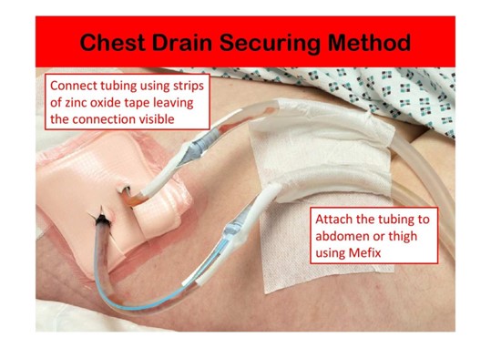
  8. Tape in place using the omental method (pictured).
  9. The drain should be attached to an underwater seal device and position confirmed with swinging and bubbling.

Chest drain securing method

Antibiotic prophylaxis

  • Administer 1.2g Co-Amoxiclav ideally before insertion and continue for 24 hours for all patients requiring a chest drain.
    • *Check Patient Allergies First*
  • Consider tetanus status for all patients.

Documentation

  • Record all procedures in medical notes.
  • Record chest drain observations in the patient notes.