Warning

All laryngectomy patients admitted to hospital should have a laryngectomy bed sign above their bed (available from ITU/ENT ward) and an appropriate cuffed trachestomy tube at their bed space.

Clinical indications for laryngectomy procedures

The clinical indications for laryngectomy procedures include:

  • carcinoma of the hypopharynx
  • carcinoma of the larynx
  • recurrence of carcinoma after radiotherapy
  • severe impairment of swallowing as a result of radionecrosis
  • obstruction of the larynx
  • recurrent aspiration pneumonia2.

Physiology

Larynx and pharynx

Reproduced with permission of סתו כסלו. This file is licensed under the Creative Commons Attribution-Share Alike 4.0 International license.

The larynx (voice box) is detached from the base of the tongue and the hypopharynx and trachea are divided. The pharynx is closed primarily and the tracheal stump is sutured to the skin to form an end tracheostomy/laryngeal stoma2. This means that the lungs have been disconnected from the mouth and nose and are only connected to the stoma in the patient’s neck. Surgical voice restoration can be performed by the creation of a tracheo-oesophageal fistula/puncture (TOF/TOP) and insertion of voice prosthesis (appendix 1). Within Ayrshire and Arran, this procedure is routinely performed at the time of surgery.

When air is inspired directly via a laryngeal stoma and not via the nasal/oral cavity, as well as loss of voice, a number of other functions are affected3:

  • Airflow resistance – airflow resistance from the upper nasal passages (nares) and trachea is important in preventing alveolar collapse and maintaining an optimum lung function. Following a laryngectomy, resistance is lost causing a decline in lung function and oxygen saturation.
  • Nasal function – inhaled air is no longer filtered, warmed or humidified by the mucous membrane of the nose and an increase in mucus production is common. Anosmia (lack of smell) will be experienced due to loss of airflow through the upper airway – this means that the patient may not smell a danger signal such as gas or fumes or food which has gone off.  Patients will also be unable to blow their nose or sniff.
  • Sphincter action of the larynx – for activities such as weight lifting, defaecation and micturition, an increase in intra-abdominal pressure is necessary and this is achieved by closure of the vocal cords.  In the absence of a larynx, patients will be unable to achieve necessary pressures for these functions. 

Communication

As a result of a total laryngectomy it is no longer possible to produce voice in the normal way4. There is subsequently a total loss of voice in the absence of a larynx and vocal folds. It is therefore necessary for the patient to learn a new method of speaking. The speech and language therapist is involved in the process of voice rehabilitation.

Differences between a laryngectomy and tracheostomy (Appendix 2)

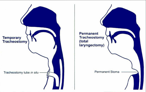
Laryngectomy - the larynx is removed and the trachea is stitched to the neck to form a permanent end stoma5. The cartilage rings of the trachea make the stoma very stable and a laryngectomy tube or tracheostomy tube may be required in the immediate post-operative period only.

Tracheostomy - a surgical incision is made and a tract is formed which must be kept open by a tracheostomy tube to maintain a patent airway. The larynx remains intact and it may be possible to voice depending on the tracheostomy tube utilised and the individuals’ medical condition.


Pre-operative preparation

  • Patients should have their physical, social and psychological needs evaluated prior to surgery.
  • The planned procedure and any questions and anxieties will be discussed with the patient and carers. Pre-operative preparation helps to reduce anxiety and patients are likely to recover better and be more satisfied if they are well informed and have had an opportunity to discuss any concerns6.
  • District nursing staff should be contacted pre-operatively to allow a pre-operative visit to be organised and baseline assessment of potential care needs can be performed.
  • The patient is given pre-operative counselling by the Macmillan Head and Neck Cancer Nurse Specialist and the Specialist Speech and Language Therapist (SLT) regarding the procedure. This includes immediate care, ongoing care and voice rehabilitation. Family and carers are involved in pre-operative counselling as a laryngectomy causes severe stress for carers7.
  • The patient should meet the physiotherapist and dietitian pre-operatively to discuss care provision.
  • Patients are given verbal information and are offered written information and a visit from a laryngectomy patient as this is known to assist coping and strongly predicts later quality of life6.
  • Smoking cessation should be discussed with relevant patients and they should be counselled to stop smoking pre-operatively if possible.

Psychological care

Explain to the patient they may experience or require some or all of the following:

  • In the immediate post-operative period, communication will be maintained by using signboards, note pads or lip reading.
  • Be unable to speak until speech rehabilitation has commenced following clinical assessment to ensure healing has occurred (day 10 post-op).
  • Require humidified oxygen therapy until stable.
  • Require tracheal suctioning to remove secretions until they are able to expectorate independently.
  • Taste and smell will be affected.
  • The patient will be unable to strain to defaecate or micturate and will be unable to lift heavy items.

Post-operative care

Post-operative bed space environment

  • The patient will be nursed in the side-room within ENT ward for at least 24 hours or the Intensive Care Unit for ventilation and to allow close monitoring of vital signs and wound site. Laminated laryngectomy bed sign should be placed above bed and a cuffed tracheostomy tube should be at the bed side.
  • Patient will have nurse call system and appropriate communication system at all times.
  • Suction equipment must be present at the bed space and operational status checked each shift.
  • Ample supply of appropriate sized sterile suction catheters must be at each bed space.
  • Alcohol based hand rub.
  • Box of non-sterile medical examination gloves (not polythene).
  • Bottle of sterile water to clean suction tubing.
  • Humidified oxygen should be available.

Post operative nursing care (day 0-10)

  • Observe closely for signs of respiratory distress and bleeding. Tracheal secretions are usually blood stained for 48 hours after surgery. Document and advise medical staff immediately if bleeding increases.
  • Chest physiotherapy will be performed as required following assessment.
  • Suction will be provided during this time as required depending on the secretions and the patient’s ability to expectorate. Suction should not be performed routinely.
  • Stoma care should be performed as required depending on secretions but should be done a minimum of twice a day. Observe wound for inflammation, swelling, exudate or wound breakdown.
  • Humidified oxygen must be administered as per protocol.
  • Nebulised saline will be administered 4 hourly as required for tenacious secretions which are difficult to expectorate.
  • Neck drains (when applicable) will be removed approximately 24-72 hours post-operatively following medical review, depending on drainage levels.
  • Stoma sutures will be removed after 7-10 days.
  • The patient will be fed by a nasogastric tube following the patient’s feeding protocol. If no leakage is evident clinically after 7-10 days then the patient will be commenced on fluid and staged diet as indicated by the speech and language therapist.

Oxygen therapy protocol

  • All laryngectomy patients who require oxygen must have oxygen therapy humidified and administered in accordance with their prescription.
  • If the patient has a laryngeal stoma (which does not require a tube) or an uncuffed tracheostomy tube in-situ then oxygen may be administered using a tracheostomy oxygen mask.
  • Ensure the weight of the oxygen tubing is supported and that the mask or T-piece has no undue tension put on it as this can lead to tube displacement and/or occlusion.
  • Ayr Hospital – tracheostomy oxygen masks are available from ITU on request. These will only be issued to patients with a laryngeal stoma or where an uncuffed tracheostomy tube is in-situ. Tracheostomy oxygen masks are available from ENT ward at Crosshouse Hospital or from Supplies Department.
  • If a laryngectomy patient requires oxygen therapy in the community then the supply of oxygen will be organised by the GP following discussion with the medical team.

Suction protocol

Suctioning protocol

Suction should be performed as required and not routinely. Patients usually only require suction in the immediate post-op period (day 1-3) and thereafter they should be encouraged to expectorate independently.   

Equipment

  • non-sterile medical exam gloves (not polythene)
  • apron
  • face mask or full face visor
  • water:
    • hospital: sterile water
    • community: cooled boiled water
  • suction tubing
  • suction catheters
  • alcohol-based hand rub
  • bowl
  • clinical waste bag
  • suction machine.

Suction catheter size:

Tube size Suction cath size
7.0mm 10FG
8.0mm 12FG
8.5mm 14FG
9.0mm 14FG
10mm 14FG

Although some charts state that size 16 and 18 catheters can be used for 9mm and 10mm tubes respectively, it is strongly recommended that the largest catheter used is a size 14 FG.

Procedure

  1. Explain procedure to patient and prepare patient.
  2. Catheter size will be selected: the diameter of the suction catheter must be less than half the inner diameter of the tracheostomy tube, to prevent hypoxia.
  3. Prepare equipment: suction tubing will be attached to suction machine, suction catheter will be placed into suction tubing, keeping the catheter wrapper in-situ to allow the end of the catheter to remain sterile. Plug in the machine to the mains supply. Pour water into the bowl.
  4. Decontaminate hands as per the World Health Organisation 5 moments for hand hygiene and HPS National Infection Prevention and Control Manual, and don personal protective equipment including mask or full face visor where there is risk of spraying or body fluids. FFP3 masks should be used for aerosol generating procedures when patient is known or suspected to have an infection such as group A strep.     
  5. Remove wrapping from suction catheter and loop catheter to prevent contamination.
  6. Switch on suction machine, ensuring pressure is below 120mmHg (16 kPa) for an adult.
  7. No suction will be applied when suction catheter is introduced into trachea/tracheostomy. If the patient is able to cough secretions as far as the end of the tracheostomy tube, then the catheter need only be inserted just beyond the end of the tube. Where the patient is unable to cough the catheter should be inserted 10-15cm.
  8. Apply suction, i.e. close over hole on Y junction on suction catheter with thumb.
  9. Withdraw catheter either in a circular motion or a side to side action.
  10. Suction should not be carried out for longer than 10 seconds.
  11. Remove catheter.
  12. Rinse suction catheter with water.
  13. Assess airway – if further suction is required repeat steps 3 to 9 using a new suction catheter (hospital) until airway is clear and breathing is less “rattly”. Within the patient’s home a suction catheter may be repassed within the same episode of care however the catheter must not be stored and re-used at a later stage.
  14. Flush suction tubing with water to remove any secretions present.
  15. Remove gloves, apron and suction catheter and discard in a disposal bag.
  16. Switch off suction machine.
  17. Perform hand hygiene.
  18. Ensure patient is comfortable.
  19. Prepare equipment, i.e. leave new catheter next to suction machine and keep wrappings intact to maintain sterility.
  20. Document relevant points into the patient’s nursing notes.

Humidification

Inhaling cold, dry air into the trachea can cause crusting of tracheal secretions. This may cause the stoma to block and therefore constant humidification is essential in the long-term management of the stoma.  Dehydration of the mucous membranes of the stoma increases the likelihood of developing an infection.  There are various types of humidification systems available and which one is used depends on the individual’s preference.

  • Buchanan protectors/laryngofoams - filter, warm and humidifying the air to ensure the laryngectomee is inhaling moistened filtered air. It is no longer necessary to wet these products as they are made of a special type of foam (hydrolox) which acts as a filter and a heat/moisture exchanger. These items are single use only in hospital however in the community some protectors may be washed following manufacturer’s instructions.
  • Heat and Moisture Exchange Systems (HMEs) - HME systems are encouraged as they provide an air tight seal for maximum filtration, humidification and improve lung resistance/function. NHSA&A currently use Provox Life Sensitive baseplates (oval/round/large) and Provox Life ‘Home’ HMEs as standard post-surgery. The specialist speech and language therapist will assess patients individually for selection of appropriate products if needed. Provox (ATOS medical) have a range of products which include baseplates, laryngectomy tubes and HME cassette filters (appendix 3). HME systems available e.g. Provox (Platon medical) have a range of products which include baseplates, laryngectomy tubes and cassette filters (appendix 3).
  • Nebulisers – assessment of the patient’s secretions should be performed, as nebulisation is not always required where adequate humidification is utilised. However, a laryngectomee who has difficulty expectorating and whose secretions are viscous should use saline nebulisers as required.  Nebuliser chambers should be washed in sterile water (hospital) or warm soapy water (community) and dried and renewed according to manufacturers’ recommendations.

Most laryngectomees will not require tracheal suction to maintain a clear airway. However, where this is indicated, equipment should be organised and patients and carers must be taught how to carry out this procedure prior to discharge.

Smoky, dusty atmospheres should be avoided initially, due to the body’s natural filtering mechanism being by-passed. Patients should also avoid using aerosol sprays. Patient/carer will be taught how to cleanse the skin around the stoma, as discussed in the stoma care section prior to discharge.

Care must be taken when bathing to avoid water entering the stoma. Patients should have their first shower, prior to discharge, with staff present to supervise. 

Patients will be advised to avoid wearing a collar that is too tight, and that shirts, sweaters etc. should be of 100% cotton/breathable material. It is advisable to wear a protector over the stoma to provide humidification and prevent soiling of the collar by tracheal secretions.

Stoma care

The aim of stoma care is to maintain a patent airway, promote healing and prevent peristomal infection, tracheitis and secondary chest infection.

  1. Perform hand hygiene before clean/aseptic task.
  2. Carers and nursing staff should wear non-sterile medical examination gloves (not polythene).
  3. Stoma care is a clean procedure. Clean around stoma area. Never use cotton wool or woven gauze swabs around the stoma as fibres may be shed into the trachea.
  4. Hospital – normal saline and non-woven gauze swabs should be used.
  5. Community – tap water and non-woven gauze swabs.
  6. If skin around the stoma is red and inflammed, a swab for microbiological investigation should be obtained and a barrier cream for example Cavilon should be applied.
  7. Crusts can be removed using forceps or tweezers.
  8. Perform hand hygiene.

Stoma buttons / lary tubes

Laryngectomees may need to wear a stoma button / lary tube to prevent reduction in size of the formed stoma.  These should be worn if instructed by the ENT consultant/staff. The patient should be able to demonstrate independence with care of the stoma button / lary tube prior to discharge. Ideally, the diameter of the stoma should be no less than 10mm. Contact ENT ward or specialist SLT for advice if the stoma measures <10mm. In some circumstances, a stoma stretch may be required. The stoma may be measured using a disposable wound ruler and size should be matched to the appropriate button/ tube size (external diameter). The patient should have a spare stoma button lary tube the same size and the stoma button / lary tube should be replaced when there is evidence of wear and tear.

Insertion of stoma button

  1. Perform hand hygiene before clean/aseptic task.
  2. Carers and nursing staff should wear non-sterile medical examination gloves (not polythene).
  3. A water-based lubricant may be used to moisten the button making insertion easier. To insert the stoma button / lary tube into the stoma compress the stoma button between the thumb and index finger, insert one edge into the stoma followed by the other edge and release pressure by the fingers allowing the button to open fully within the stoma. Ensure inner flange is within the trachea.
  4. Ensure button / lary tube is tight within the stoma and will not be easily expelled/inhaled. A smaller size button than the one the patient normally wears should be given to the patient where possible (smallest size 10.5mm external diameter). This can be inserted if there is difficulty inserting the original button / lary tube.
  5. A thread/string may be looped through the hole on the outer flange of the button/ lary tube: the thread is then attached to the patients clothing. This prevents the button / lary tube being lost/expelled.
  6. Perform hand hygiene.

To remove stoma button

  1. Loosen attached thread from clothing (if used).
  2. Gently pull on stoma button / lary tube and ease from trachea.
  3. Wash stoma button with sterile water (hospital) or tap water (community) and allow to air dry.

Voice rehabilitation

There are three methods of voice rehabilitation available to the laryngectomy patient4:

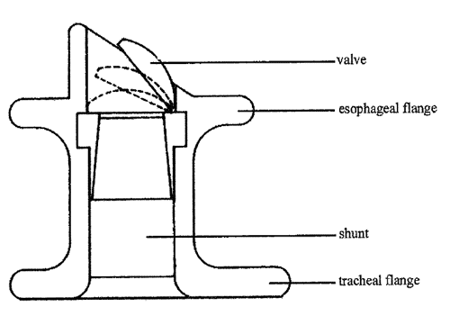
  • Surgical voice restoration (SVR) – SVR is the most frequently used method of voice restoration after a total laryngectomy. The voice prosthesis can be inserted at the time of surgery into a surgically created fistula between the trachea and oesophagus (appendix 4). When the stoma is occluded the voice prosthesis allows exhaled air from the lungs to enter the oesophagus and sound is generated.  The voice prosthesis itself does not generate any voice or sound but only allows air from the lungs to enter the oesophagus. The vibration is then modified into speech by the lips, tongue and teeth (appendix 5).
  • Oesophageal speech – this is an older method of voice rehabilitation (swallow air and ‘burp’ back). The patient has to be taught how to force air into the oesophagus from the mouth to cause vibration for voice. The vibration is modified by the lips, tongue and teeth to form speech.
  • Artificial (electronic) larynx – the artificial larynx is a hand-held mechanical device. The device is held against the neck and when switched on vibrations are transmitted through the tissues. The vibrations are then modified by the lips, tongue and teeth.

Voice quality with voice prosthesis

There are two manufacturers of voice prosthesis globally for laryngectomy SVR – Provox and Blom-Singer. Both companies make a range of valves to suit different sizes / shapes of fistula and to address voicing issues. Within Ayrshire and Arran, the Provox Vega voice prosthesis is routinely utilised for voice rehabilitation following total laryngectomy (appendix 5).

Voice quality may temporarily deteriorate when a patient first learns to speak with a valve.

  • Encourage the patient to relax.
  • Ensure the valve is clean and there is no leakage.
  • Ensure the patient occludes the stoma when talking.

Care of Provox valve

Care of the Provox voice prosthesis

For the care of other voice prosthesis please refer to the manufacturer’s instructions or contact Head & Neck Speech & Language Therapy Team / CNS or ENT ward for advice.

How long does the Provox valve last?

The prosthesis is not a permanent implant and needs periodic replacement. Both manufacturers advise a valve should last 3-4 months. The primary indication for replacement is incompetence of the valve due to candida causing leakage of fluids through the middle of the prosthesis.

How to check for leakage/incompetence of the valve/leakage through middle of valve

  • Ask patient to take a drink of a visible liquid i.e. milk.
  • Use a pen torch and observe for leakage through the middle of the valve.
  • If leakage is observed contact the Head & Neck Speech & Language Therapy Team for an appointment for the valve to be replaced.
  • Use the valve plug when eating and drinking until the valve has been changed. The valve plug can be removed after eating and drinking to allow the patient to speak as normal. A valve plug will be provided prior to discharge.

Leakage around valve

  • Ask patient to take a drink of a visible liquid.
  • Use a pen torch and observe for leakage around the valve.
  • If leakage is visible contact the Head & Neck Speech & Language Therapy Team for an appointment for valve to be replaced.
  • Valve change clinics currently operate within Out-Patient Waiting Area 1 on Friday afternoons. An appointment can be made to have the valve assessed and changed by contacting the Head & Neck Speech & Language Therapy Team.
  • Patient may have to be admitted to ENT ward for management. The valve may require to be removed to allow the fistula to shrink before re-insertion.

Cleaning the Provox valve

Equipment required:

  • PPE
  • Provox valve brush
  • Small bowl with sterile water (hospital)/tap water (community)
  • Non-woven gauze swabs
  • Pen torch.

To keep the Provox valve working properly it should be cleaned twice a day, morning and night. Cleaning may have to be performed more regularly when secretions are increased e.g during chest infections.

Procedure 8:

  1. Explain the procedure and prepare the patient: place in an upright position if possible.
  2. Prepare equipment and area where the care will be performed.
  3. Decontaminate hands as per the World Health Organisation 5 moments for hand hygiene and HPS National Infection Prevention and Control Manual.
  4. Nursing staff should wear PPE including face mask/visor where there is a risk of splashing of spraying of body fluids. FFP3 masks should be worn for aerosol generating procedures when patient known or suspected as having an infection such as group A strep.
  5. Identify the valve (a pen torch may be required if the lighting is poor).
  6. The Provox brush can be used dry or it can be wet with sterile water (hospital) or tap water (community) and shake off any excess water.
  7. Insert the Provox brush until all the bristles are inside the valve - the blue collar on the brush prevents the brush being inserted any further. Rotate the brush 90o to the left then the right, with a gentle ‘twisting movement’.
  8. Remove brush and clean with a gauze swab or by rinsing with water.
  9. Repeat steps 6 - 8 until the valve is clear (i.e. until no debris is removed from the valve).
  10. The valve may rotate when cleaning. The valve will not come out of the fistula.
  11. When cleaning is complete, make sure ‘tail’ (dimple) is in the correct position for optimum voice production, i.e. at the bottom – 6 o’clock position.
  12. Once the prosthesis is clear, clean the brush by washing it in sterile water (hospital) or warm soapy water (community), then rinse and dry thoroughly.
  13. Ensure the patient is comfortable.
  14. Perform hand hygiene.
  15. If the patient is away from home, they may clear the valve by occluding the stoma and giving a hard cough and this may clear the blockage. The laryngectomee should carry a Provox brush for cleaning at all times.
  16. Document care given.
  17. The Provox brush should be disposed of when it shows signs of wear and tear (as per manufacturers’ instructions) and replaced with a new brush.

Factors which influence the quality of voice:

  • Encourage patient to follow any advice given by the Specialist SLT.
  • Reassure patient if anxious.
  • Encourage relaxed posture and avoid neck/shoulder muscle tension.
  • Complete occlusion of the stoma is required during voicing.

Emergency situations

Blockage of stoma/trachea

  • Remove obstruction with forceps if it is visible.
  • Humidify with nebulised saline to help loosen secretions.
  • Where available (e.g. paramedic services/hospital setting) apply suction in an attempt to remove obstruction.
  • Community - if unable to remove the obstruction dial 999 for emergency assistance.
  • If this is an ongoing problem, contact ENT team for advice or for patient to be reviewed.

Haemorrhage

  • Assess amount of bleeding. Small amounts of blood stained secretions can be expected.
  • Within the community if blood loss is more than streaking, the patient should be reviewed by a GP or in case of excessive bleeding, emergency assistance should be sought (dial 999).
  • Where available (e.g. paramedic services/hospital setting) a cuffed tracheostomy tube will be inserted and the cuff inflated. This will apply pressure, which may help to control bleeding and will prevent the aspiration of blood coming from any point above the cuff. Urgent review by ENT medical team required.

Resuscitation of a patient with a laryngectomy

National Tracheostomy Safety Project. Emergency laryngectomy management

Hospital setting

Patients who have undergone laryngectomy have no upper airway so mouth to mouth resuscitation and endotracheal intubation is not possible. Access to the airway is only possible via the neck stoma. In the event of a respiratory or cardiac arrest artificial ventilation must be given via the laryngeal (neck) stoma.  Any crusting or obstruction should be removed from the stoma with forceps. A cuffed tracheostomy tube should be inserted and the tracheostomy cuff should be inflated immediately to provide ventilation. Where a cuffed tracheostomy tube is not available the patient should be ventilated using a paediatric face mask over stoma.  In the hospital setting all patients admitted with a laryngectomy must have an appropriate size of cuffed tracheostomy tube readily available at the bed space.

  • Ventilate via paediatric face mask over stoma or cuffed tracheostomy tube with cuff inflated.
  • The tracheostomy tube is connected to the ambubag via a catheter mount and the patient is ventilated manually using oxygen at 10litres/min.
  • Nose and mouth should be occluded throughout CPR to prevent air leakage from the tracheo-oesophageal fistula.
  • CPR should be performed via the laryngeal stoma as per NHS Ayrshire and Arran guidelines.

Community setting

  • Call an ambulance and summon medical help.
  • Lie patient flat.
  • Any crusting or obstruction should be removed from the stoma with forceps.
  • Nose and mouth should be occluded throughout CPR to prevent air leakage from the tracheo-oesophageal fistula.
  • CPR via laryngeal stoma will continue until ambulance/medical assistance arrives.

Self care and discharge planning

Self care

The patient should be taught and encouraged to become independent with all aspects of laryngectomy and stoma care at the earliest opportunity post-operatively and education should be ongoing3. This process should be integrated into the patient’s normal activities of daily living.

Psychological impact of the laryngectomy

Laryngectomy results in loss of ‘normal’ voice and formation of a stoma. These changes mark the individual as different and some stigma may be attached to the changes causing negative reactions in others9.  Perceived stigma can have a significant impact on work, recreation, financial situation, family relationships, social relationships, community involvement and self-expression and result in low mood and depression.  Nurses can plan care strategies to minimise perceived and actual stigmatization and promote adjustment to the laryngectomy by the patient and their carers/family9.

Discharge planning

The discharge planning of any patient should commence prior to or on admission to hospital of a patient undergoing major head and neck surgery. Visiting the patient at home, prior to surgery would permit the community nurse to commence an assessment of the patient’s needs and social situation while the communication difficulties are limited. This may identify any potential issues or problems.

Discharge planning meetings should be encouraged, as this facilitates a multi professional approach to discharge and enables a seamless continuation of care.

If any relevant equipment is required to promote the discharge this will be the ideal opportunity for the community nurse to assess and plan for discharge. Within Ayrshire and Arran equipment is readily available for rapid delivery.

Once home the community nurse should be able to visit (if previously unable to) to asses both patient and home circumstances and any difficulties would be addressed. Community nurses work very closely with Social Services Departments and are able to liaise with colleagues to support patients at home. During this initial visit the community nurse would be aware of the patient’s level of independence with stoma care and of their present speech ability at this time. The community nurse would also be able to prescribe and or order any supplies the patient requires to continue with their stoma care. Community nurses will also be able to liaise with GP and community pharmacist with regards to medications required.

The nurse and the patient should be able to agree time and dates for further support contact. At this point the community nurse would give the patient the contact telephone numbers for the both the community nurses base and the out of hours contact details.

Training and education

Community nursing staff are encouraged to attend ENT ward to go over care prior to discharge of a patient with a laryngectomy. Alternatively, a joint visit may be performed with the Macmillan Head and Neck Cancer Nurse Specialist following discharge.

Individual patient care can be discussed and advice can be obtained by contacting ENT ward, Specialist SLT, Macmillan Head and Neck Cancer Nurse Specialist or ENT Nurse Practitioner.

Equality and diversity impact assessment

This guideline has been Impact Assessed using the NHS Ayrshire and Arran Equality and Diversity Impact Assessment Toolkit.      

Staff are reminded that they may have patients who require communication in a form other than English e.g. other languages or signing. Additionally, some patients may have difficulties with written material.

At all times, communication and material should be in the patients preferred format. This may also apply to patients with learning difficulties.

In some circumstances there may be religious and/or cultural issues which may impact on this guideline e.g. choice of gender of health care professional. Consideration should be given to these issues when treating/examining patients.

References

Related NHS Ayrshire & Arran and HPS documents

Health Protection Scotland (2015) National Infection Prevention and Control Manual.

NHS Ayrshire and Arran (2013) Reference guide for the management of an adult with a tracheostomy (hospital and community setting)

References 

  1. Markovitch H (2006) Blacks Medical Dictionary 41st Edition Scarecrow Press London.
  1. Buckley G (2000) Surgical management at specific sites Chapter 4.2 in Feber T (2000) Head and Neck Oncology Nursing Whurr Publishers Ltd London p434-443.
  1. Feber T (2000) Airway Management Chapter 2.1 in Feber T (2000) Head and Neck Oncology Nursing Whurr Publishers Ltd London p89-120.
  1. Greene M and Mathieson L (2001) The Voice and Its Disorders 6th Edition Whurr Publishers London p597.
  1. Buckley G (2000) Surgical management at specific sites Chapter 4.2 in Feber T (2000) Head and Neck Oncology Nursing Whurr Publishers Ltd London p434-442.
  1. Stam HJ, Koopman’s JP and Mathieson CM (1991) The psychological impact of laryngectomy: a comprehensive assessment Journal of Psychological Oncology 9 (3) p37-59.
  1. Krouse HJ, Rudy SF, Vallerand AH, Hickey MM, Klein MN, Kagan SH & Walizer EM (2004) Impact of tracheostomy or laryngectomy on spousal and caregiver relationships Head and Neck Nursing 22 (1) p10-25.
  1. Platon Medical (2001) Looking after your Provox Voice Prosthesis Platon Medical London.
  1. Feber T (1998) Design and evaluation of a strategy to provide support and information for people with cancer of the larynx European Journal of Oncology Nursing 2 (2) p106-114.
  1. NHS Ayrshire and Arran (2007) Manual of Infection Control.
  1. National Cancer Institute (2015) Hypopharyngeal Cancer Treatment. Available from: http://www.cancer.gov/types/head-and-neck/patient/hypopharyngeal-treatment-pdq (Accessed: 26.02.16)

Appendix 1: Pre and post laryngectomy anatomy showing tracheoesophageal puncture/fistula and speaking valve

The larynx is removed and the trachea is separated from the oesophagus and stitched onto the neck. This means that the nose and mouth is no longer attached to the trachea and therefore all breathing is accomplished through the laryngeal stoma and the humidification and filtration functions of the nose are lost.

Photograph of a patient following a laryngectomy - stoma and speaking valve indicated:-

Photograph with a larybutton inserted to keep stoma size stable - a filter may be added to filter and humidify air and increase airway resistance:-

Please note - picture shows larybutton not stoma stud

Appendix 2: Differences in anatomy between temporary tracheostomy and permanent tracheostomy (laryngectomy procedure)

The larynx is removed and the trachea is stitched to the neck to form a permanent end stoma. The cartilage rings of the trachea make the stoma very stable.

Appendix 3: Humidification and moisture exchange systems (HME)

An HME system humidifies and filters the inhaled air and increases lung resistance. When the patient covers over the HME on exhalation, air is forced through the one way speaking valve into the oesophagus to produce vibration at the neoglottis (pharyngo-esophageal (PE) segment). This produces sound which is modified by the lips, tongue and teeth to form speech.

Appendix 4: Speaking valve

When the laryngeal stoma is covered on exhalation, air is forced through the one way speaking valve into the oesophagus to produce vibration at the neoglottis (pharyngo-esophageal (PE) segment). This produces sound which is modified by the lips, tongue and teeth to form speech. The one-way mechanism prevents fluid and diet from leaking back into the trachea and lungs as the valve closes following exhalation. If fluid and diet leak through the valve then this indicates that the valve needs to be reviewed and replaced.

Appendix 5: Voice production

By covering the laryngeal stoma on exhalation, exhaled air is directed into the oesophagus and the vibration is then modified by the lips, tongue and teeth to produce speech.

Appendix 6: Product ordering information

Product Order information
Buchanan protector

Stores or prescription

Order no: LABUP 0001

ATOS Medical

Buchanan lite bibs (large)

Pack of 10

Prescription

Order no: LABLT 1001 (white), LABLT 1002 (blue), LABLT 1003 (beige)

ATOS Medical

Laryngofoam (large)

Pack of 30

Stores or prescription

Order no: LALFF0023

ATOS Medical

Provox brushes

(Pack of 6)

Prescription

Order no:8404

ATOS Medical

Provox plug

Ref: 7205

20Fr valve order ref: 8129

ATOS Medical

Shower shield

Non-stock order

Order no: LASHC0001

ATOS Medical

Shower aid

For use with baseplate (in place of HME) for showering

Non-stock order

Order no: 7260

ATOS Medical

Mirror, pen torch, plastic tweezers, radar key and care kit bag

Provided free of charge by laryngectomy home delivery companies. 

Supply of bags/kit on ENT ward and in UHC Speech Therapy Department.

Editorial Information

Last reviewed: 09/03/2023

Next review date: 17/04/2026

Author(s): McCallum C, Rennie C.

Version: 05.0

Author email(s): claire-ann.mccallum@aa.nhs.scot, caroline.rennie@aa.nhs.scot.

Approved By: Boyd D

Reviewer name(s): Boyd D, Head and Neck Governance Group.

Internal URL: http://athena/cgrmrd/ClinGov/DraftGuidance/G008%20Guideline%20for%20Laryngectomy%20Care.pdf