  1. Right Decisions
  2. Maternity & Gynaecology Guidelines
  3. Maternity
  4. Back
  5. Ultrasound
  6. Mild Ventriculomegaly on Antenatal Ultrasound (916)
Important: please update your RDS app to version 4.7.3 Details with newsletter below.

Please update your RDS app to v4.7.3

We asked you in January to update to v4.7.2.  After the deployment planned for 27th February, this new update will be needed to ensure that you are able to download RDS toolkits even when the RDS website is not available. We will wait until as many users as possible have downloaded the new version before switching off the old system for app downloads and moving entirely to the new approach.

To check your current RDS version, click on the three dots bottom right of the RDS app screen. This takes you to a “More” page where you will see the version number. 

To update to the latest release:

 On iPhones – go to the Apple store, click on your profile icon top right, scroll down to see the apps waiting to be updated and update the RDS app.

On Android phones – these can vary, but try going to the Google Play store, click on your profile icon top right, click on “Manage apps and device”, select and update the RDS app.

Right Decision Service newsletter: February 2025

Welcome to the February 2025 update from the RDS team

1.     Next release of RDS


A new release of RDS is planned (subject to outcomes of current testing) for week beginning 24th February. This will deliver:


  • Fixes to mitigate the recurring glitches with the RDS admin area and the occasional brief user interface outages which have arisen following implementation of the new distributed technology infrastructure in December 2024.


  • Capability to embed content from Google calendar, Google Maps, Daily Motion, Twitter feeds, Microsoft Stream into RDS pages.


  • Capability to include simple multiplication in RDS calculators.


The release will also incorporate a number of small fixes, including:

  • Exporting of form within Medicines Sick Day Guidance in polypharmacy toolkit
  • Links to redundant content appearing in search in some RDS toolkits
  • Inclusion of accordion headers alongside accordion text in search result snippets.
  • Feedback form on mobile app.
  • Internal links on mobile app version of benzo tapering tool


We will let you know when the date and time for the new release are confirmed.


2.     New RDS developments

There is now the capability to publish toolkits on the web with left hand side navigation rather than tiles on the homepage. To use this feature, turn on the “Toggle navigation panel” option at the top of the Page settings menu at toolkit homepage level – see below. Please note that publication to downloadable mobile app for this type of navigation is still under development.

The Benzodiazepine tapering tool (https://rightdecisions.scot.nhs.uk/benzotapering) is now available as part of the RDS toolkit for the national benzodiazepine prescribing guidance developed by the Scottish Government Effective Prescribing team. The tool uses this national guidance developed with a wide-ranging multidisciplinary group. This should be used in combination with professional judgement and an understanding of the needs of the individual patient.

3.     Archiving and version control and new RDS Search and Browse interface

Due to the intensive work Tactuum has had to undertake on the new technology infrastructure has pushed back the delivery dates again and some new requirements have come out of the recent user acceptance testing. It now looks likely to be an April release for the search and browse interface. The archiving and version control functionality may be released earlier. We’ll keep you posted.

4.     Statistics

At the end of January, Olivia completed the generation of the latest set of usage statistics for all RDS toolkits. If you would like a copy of the stats for your toolkit, please contact Olivia.graham@nhs.scot .


5.     Review of content past its review date

We have now generated reports of all RDS toolkit content that has exceeded its review date by 6 months or more. We will be in touch later this month with toolkit owners and editors to agree the plan for updating or withdrawing out of date content.


6.     Toolkits in development

Some important toolkits in development by the RDS team include:

  • National CVD prevention pathways – due for release end of March 2025.
  • National respiratory pathways, optimal cancer diagnostic pathways and cancer prehabilitation pathways from the Centre for Sustainable Delivery. We will shortly start work on the national cancer referral pathways, first version due for release via RDS around end of June 2025.
  • HIS Quality of Care Review toolkit – currently in final stages of quality assurance.


The RDS team and other information scientists in HIS have also been producing evidence summaries for the Scottish Government Realistic Medicine team, to inform development of national guidance around Procedures of Limited Clinical Value. This guidance will in due course be translated into an RDS toolkit.


7. Training sessions for new editors (also serve as refresher sessions for existing editors) will take place on the following dates:

  • Friday 28th February 12-1 pm
  • Tuesday 11th March 4-5 pm


To book a place, please contact Olivia.graham@nhs.scot, providing your name, organisation, job role, and level of experience with RDS editing (none, a little, moderate, extensive.)


To invite colleagues to sign up to receive this newsletter, please signpost them to the registration form  - also available in End-user and Provider sections of the RDS Learning and Support area.   If you have any questions about the content of this newsletter, please contact his.decisionsupport@nhs.scot  If you would prefer not to receive future newsletters, please email Olivia.graham@nhs.scot and ask to be removed from the circulation list.

With kind regards


Right Decision Service team

Healthcare Improvement Scotland


Mild Ventriculomegaly on Antenatal Ultrasound (916)

Warning
Please report any inaccuracies or issues with this guideline using our online form

Fetal ventriculomegaly is a common finding on antenatal ultrasound and is defined as an atrial measurement of ≥ 10mm of the posterior horn of the lateral ventricle (1). It can be further subdivided into mild 10-12mm, moderate 13- 15mm and severe >15mm (2). It has a prevalence of approximately 1% (3). Ventriculomegaly has a range of causes; normal variation, aneuploidy, genetic syndromes, primary brain structural abnormalities, congenital infections, cerebrovascular accidents and intracranial haemorrhage. Prognosis and corresponding counselling of the parents is dependent on the cause of the ventriculomegaly, the antenatal progression and any co-existing abnormalities(4). It is therefore vitally important to look for  any underlying aetiologies and co-existing CNS and non-CNS abnormalities in order to present the parents with the most relevant and accurate information.

Diagnosis

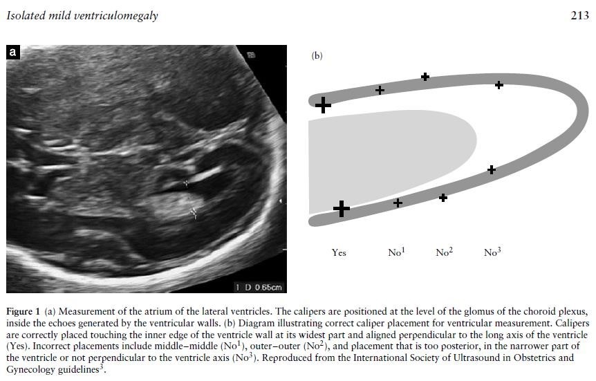
Accurate measurement of the ventricles is important in both defining ventriculomegaly and also assessing progression. The fetal head should be scanned in the axial plane at the level of the frontal horns and the cavum septum pellucidum (CSP) (the same level at which a head circumference is taken), at an appropriate magnification that the head fills the screen. The callipers should be placed at the internal margins of the atrial walls at the level  of the parietal occipital groove and the glomus of the choroid plexus, perpendicular to the axis of the ventricle.

Although the distal ventricle is always easier to see than the proximal one because of reflection of the ultrasound beams from the fetal skull, both ventricles should be checked; ventriculomegaly is unilateral in 50-60% cases and bilateral in 40-50% (5).

Ultrasonography

Once ventriculomegaly has been diagnosed, there should be a detailed, sonographic evaluation of the neuroanatomy by a medical sonographer. Whether this is by transabdominal or transvaginal ultrasound will depend on the preference of the patient, the sonographer and the fetal position.

Other, non-CNS structures should also be carefully assessed including fetal biometry looking for evidence of growth restriction, the heart and any markers of intrauterine infection.

Testing for genetic disorders

Parents should be offered invasive, diagnostic testing and chromosomal microarray (CMA). 

Between 0-5% (2, 5) of fetuses with apparently isolated m i l d ventriculomegaly will have an underlying abnormal karyotype, most commonly Trisomy 21 and a further 10-15% will have abnormalities found on CMA.  

Testing for fetal infection

Congenital infections, most commonly cytomegalovirus (CMV), toxoplasmosis, parvovirus and Zika have been associated with mild ventriculomegaly in around 8% of cases (5). Parents should be offered tests for CMV, toxoplasmosis and parvo virus (regardless of history of known exposure or symptoms). Women with mild fetal ventriculomegaly who have been to a Zika area and not yet tested should be offered a test.

Fetal MRI

Fetal MRI (fMRI) can be a useful adjunct to ultrasound if the relevant radiological expertise and technology is available. The additional information will depend on the size of the ventricles as well as the quality of the original ultrasound and the level of expertise in the practitioner. The chance that fMRI will find important, clinically relevant additional brain abnormalities not picked up on ultrasound varies in the literature from 1-14% with the most recent studies putting the figure at 5-6% (5). The most common abnormality picked up on fMRI after being missed on ultrasound is agenesis of the corpus callosum.

Women wishing to have a fetal MRI, to look for additional brain abnormalities that may affect the prognosis, after appropriate counselling should be referred to fetal medicine department for review.

  • The fetal medicine department will arrange the MRI
  • Fetal medicine will review again after MRI to discuss results.
  • Thereafter the patient will go back to their own unit, unless otherwise indicated and delivery will be planned in their own unit.

Follow up antenatal ultrasound examinations

There are no data on optimal timings of follow up assessments once a diagnosis has been made. A suggested pragmatic approach would be 4-weekly assessments. Progression of ventriculomegaly is an important prognostic indicator; evidence suggests that 5% progress during pregnancy (5).

Delivery

The timing and mode of delivery should be planned as per normal obstetric indications. An alert should be placed on the electronic BadgerNet record to ensure that neonatologists are made aware of the antenatal diagnosis.

Cord bloods should be taken with parental consent for chromosomal analysis and congenital viral infections from those infants who didn’t have antenatal testing.

Postnatal follow up should be arranged by the neonatologists prior to discharge from hospital.

Prognosis

Most of the statistics quoted in the literature are based on whether the ventriculomegaly is apparently isolated or not; true isolation will only be able to be confirmed postnatally. Neurodevelopmental delay in case of isolated unilateral mild or moderate ventriculomegaly is thought to be 6% (5); in bilateral isolated ventriculomegaly this rises to 8-12% (7). This may not be dramatically higher than the background population risk. Long-term prognosis also depends on associated findings and the positive results of any investigations. 

Parents should be offered antenatal counselling by paediatricians to discuss prognosis and postnatal care in greater detail. A patient information leaflet from ISUOG and a link to further information is below.

Further information

Melchiorre, K & Bhide, Amarnath & Gika, Artemis & Pilu, G & Papageorghiou, A.T. (2009). Counseling in isolated mild ventriculomegaly. Ultrasound in Obstetrics & Gynecology

Patient Information:

ISUOG. Ventriculomegaly
"This leaflet is to help you understand what Ventriculomegaly is, what tests you need, and the implication of having been diagnosed with Ventriculomegaly for you, your baby and your family."

Editorial Information

Last reviewed: 01/04/2021

Next review date: 01/04/2024

Author(s): Rachel Bradnock.

Approved By: Obstetrics Clinical Governance Group

Document Id: 916

References
  1. International Society of Ultrasound in Obstetrics and Gynecology Education Committee. Sonographic examination of the fetal central nervous system: guidelines for performing the ‘basic examination’ and the ‘fetal neurosonogram’. Ultrasound Obstet Gynecol 2007; 29: 109 – 116.
  2. Society for Maternal-Fetal Medicine (SMFM): Fox NS et al. Mild Fetal Ventriculomegaly: diagnosis, evaluation and management. SMFM Consult Series 45 2018.
  3. Pilu G, Hobbins JC. Sonography of fetal cerebrospinal anomalies. Prenat Diagn 2002; 22: 321 – 330.
  4. Scala C, Familiari A, Pinas A, Papageorghiou T, Bhide A, Thilaganathan B, Khalil A. Perinatal and long-term outcomes in fetuses diagnosed with isolated unilateral ventriculomegaly: systematic review and meta-analysis. Ultrasound Obstet Gynecol 2017; 49: 450–459
  5. Griffiths PD, Brackley K, Bradburn M, et al. Anatomical subgroup analysis of the MERIDIAN cohort: ventriculomegaly. Ultrasound Obstet Gynecol 2017;50:736-44.
  6. RCOG/RCM/PHE/HPS Clinical Guidelines. Zika Virus Infection and Pregnancy. Updated Feb 2019.
  7. Pagani G, Thilaganathan B, Prefumo F. Neurodevelopmental outcome in isolated mild fetal ventriculomegaly: systematic review and meta-analysis. Ultrasound Obstet Gynecol 2014; 44: 254 – 260.