  1. Right Decisions
  2. Maternity & Gynaecology Guidelines
  3. Gynaecology
  4. Back
  5. Gynaecology guidelines
  6. Premenopausal Ovarian Masses (514)
Announcements and latest updates

Welcome to the Right Decision Service (RDS) newsletter for August 2024.

  1. Contingency planning for RDS outages

Following the recent RDS outages, Tactuum and the RDS team have been reviewing the learning from these incidents. We are committed to doing all we can to ensure a positive outcome by strengthening the RDS to make it fully robust and clinically resilient for the future.

We would like to invite you to a webinar on 26th September 3-4 pm on national and local contingency planning for future RDS outages.  Tactuum and the RDS team will speak about our business continuity plans and the national contingency arrangements we are putting in place. This will also be a space to share local contingency plans, ideas and existing good practice. We would also like to gather your views on who we should send communications to in the event of future outages.

I have sent a meeting request for this date to all editors – please accept or decline to indicate attendance, and please forward on to relevant contacts. You can also contact Olivia.graham@nhs.scot directly to register your interest in participating.


2.National  IV fluid prescribing  calculator

This UK CA marked calculator is now live at https://righdecisions.scot.nhs.uk/ivfluids  . It has been developed by a multiprofessional steering group of leads in IV fluids management, as part of the wider Modernising Patient Pathways Programme within the Centre for Sustainable Delivery.  It aims to address a known cause of clinical error in hospital settings, and we hope it will be especially useful to the new junior doctors who started in August.

Please do spread the word about this new calculator and get in touch with any questions.


  1. New toolkits

The following toolkits are now live;

  1. Updated guidance on current and future Medical Device Regulations

We have updated and simplified this guidance within our standard operating procedures. We have clarified the guidance on how to determine whether an RDS tool is a medical device, and have provided an interactive powerpoint slideset to steer you through the process.


  1. Guide to six stages of RDS toolkit development

We have developed a guide to support editors and toolkit leads through the process of scoping, designing, delivering, quality assuring and implementing a new RDS toolkit.  We hope this will help in project planning and in building shared understanding of responsibilities throughout the full development process.  The guide emphasises that the project does not end with launch of the new toolkit. Implementation, communication and evaluation are ongoing activities throughout the lifetime of the toolkit.


  1. Training sessions for new editors (also serve as refresher sessions for existing editors) will take place on the following dates:
  • Thursday 5 September 1-2 pm
  • Wednesday 24 September 4-5 pm
  • Friday 27 September 12-1 pm

To book a place, please contact Olivia.graham@nhs.scot, providing your name, organisation, job role, and level of experience with RDS editing (none, a little, moderate, extensive.)

7 Evaluation projects

Dr Stephen Biggart from NHS Lothian has kindly shared with us the results of a recent survey of use of the Edinburgh Royal Infirmary of Edinburgh Anaesthesia toolkit. This shows that the majority of consultants are using it weekly or monthly, mainly to access clinical protocols, with a secondary purpose being education and training purposes. They tend to find information by navigating by specialty rather than keyword searching, and had some useful recommendations for future development, such as access to quick reference guidance.

We’d really appreciate you sharing any other local evaluations of RDS in this way – it all helps to build the evidence base for impact.

If you have any questions about the content of this newsletter, please contact his.decisionsupport@nhs.scot  If you would prefer not to receive future newsletters, please email Olivia.graham@nhs.scot and ask to be removed from the circulation list.


With kind regards


Right Decision Service team

Healthcare Improvement Scotland

Premenopausal Ovarian Masses (514)

Warning

Please report any inaccuracies or issues with this guideline using our online form

This guideline has been produced to assist clinicians with the initial assessment and appropriate management of suspected benign ovarian masses in premenopausal women. Up to 10% of women will have some form of surgery during their lifetime for the presence of an ovarian mass. In pre-menopausal women almost all ovarian masses and cysts are benign. The overall incidence of a symptomatic ovarian cyst in a pre-menopausal female being malignant is approximately 1:1000, increasing to 3:1000 at the age of 50. Pre-operative differentiation between the benign and the malignant ovarian mass in the pre-menopausal woman can be problematic with no specific tests. Exceptions are germ cell tumours with elevations of specific tumour markers such as alphafetoprotein (α-FP) and human chorionic gonadotrophin (hCG).

For the purposes of this guideline, simple cysts of 3cm or less should be considered physiological and do not merit further investigation.

The aim should be to minimise patient morbidity by conservative management where possible, use of laparoscopic techniques where appropriate, and referral to the gynaecological oncologists where appropriate.

History

A thorough medical history should be taken from the woman with specific attention to risk factors or protective factors for ovarian malignancy and a family history of ovarian or breast cancer.

Symptoms suggestive of endometriosis should be specifically considered along with any symptoms suggesting possible ovarian malignancy: persistent abdominal distension, appetite change including increased satiety, pelvic or abdominal pain, increased urinary urgency and/or frequency.

Examination and Investigations

A careful physical examination of the woman is essential and should include abdominal and vaginal examination, and examination to determine the presence or absence of local lymphadenopathy. Although clinical examination has poor sensitivity in the detection of ovarian masses, its importance lies in the evaluation of mass tenderness, mobility, nodularity and ascites.

In the acute presentation with pain the diagnosis of accident to the ovarian cyst should be considered (torsion, rupture, haemorrhage).

Imaging

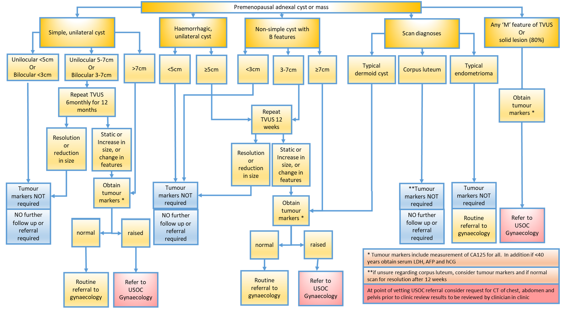
A pelvic ultrasound is the single most effective way of evaluating a pelvic mass with transvaginal ultrasonography being preferable due to its increased sensitivity over transabdominal ultrasound. Routine use of CT or MRI is not indicated but where clinical or ultrasound suspicion exists, refer to Guidelines for Imaging of Gynaecological Malignancy (West of Scotland Cancer Network Guideline). CT of the abdomen and pelvis should be performed for masses with RMI >200 or in those with RMI <200 where clinical or ultrasound suspicion exists. MRI pelvis / lower abdomen should be performed in those with a complex mass which is difficult to characterise clinically or on ultrasound, or in young women (<30yrs) with suspected malignant tumour or a complex pelvic mass.

Blood tests

  1. CA125 – a serum CA125 assay does not need to be undertaken in all premenopausal women when an ultrasonographic diagnosis of a simple ovarian cyst has been made, but should be performed in all other circumstances. However it must be recognised that it is unreliable in determining whether ovarian lesions are benign or malignant as CA125 is also raised in conditions such as fibroids, endometriosis, adenomyosis and pelvic infection. Note also that CA125 is primarily a marker for epithelial ovarian tumours but is only elevated in around 50% of early stage disease.
  2.  LDH, αFP and hCG should be measured in all women under the age of 40 with a complex ovarian mass to exclude germ cell tumours.
  3. Calculate RMI – see below.

Calculation of the RMI

RMI combines three presurgical features: serum CA125 (CA125); menopausal status (M); and ultrasound score (U).
The RMI is a product of the ultrasound scan score, the menopausal status and the serum CA125 level (IU/ml) as follows:     RMI = U x M x CA125

  • The ultrasound result is scored 1 point for each of the following characteristics: multilocular cysts, solid areas, metastases, ascites and bilateral lesions. U = 0 (for an ultrasound score of 0), U=1 (for an ultrasound score of 1, U=3 (for an ultrasound score of 2-5).
  • The menopausal status is scored as 1=premenopausal and 3=postmenopausal.
  • Postmenopausal can be defined as women who have not had a period for more than one year or women over the age of 50 who have had a hysterectomy.
  • Serum CA125 is measured in IU/ml and can vary between zero to hundreds or even thousands of units.

Management

  • Women with an RMI of more than 200 should be discussed with the gynaecological oncology team and presented to the managed clinical network for gynaecological oncology after appropriate imaging as per WOSCAN Guidelines.
  • Women with small (less than 50mm diameter) ovarian cysts generally do not require follow-up as these cysts are very likely to be physiological and almost all resolve within 3 menstrual cycles.
  • Women with simple ovarian cysts of 50-70mm in diameter should have yearly ultrasound follow-up and those with larger simple cysts should be considered for either further imaging or surgical intervention.
  • Ovarian cysts that persist or increase in size are unlikely to be functional and may warrant surgical management.
  • The use of the combined oral contraceptive pill does not promote the resolution of ovarian cysts.

Surgery

  • A laparoscopic approach should be used whenever possible.
  • Aspiration of ovarian cysts, either vaginally or laparoscopically, is less effective and is associated with a high rate of recurrence.
  • Spillage of cyst contents should be avoided where possible as pre-operative and intra-operative assessment cannot absolutely preclude malignancy.
  • Where minimal access surgery is employed, consideration should be given to the use of a tissue bag to avoid peritoneal spill of cystic contents, bearing in mind the likely pre-operative diagnosis.
  • The possibility of oophorectomy must be discussed prior to surgery, documented in the notes and included in the consent form.

Editorial Information

Last reviewed: 14/12/2016

Next review date: 30/04/2023

Author(s): Morton Hair.

Approved By: Gynaecology Clinical Governance Group

Document Id: 514

References

RCOG. Management of Suspected Ovarian Masses in Premenopausal Women (Greentop Guideline No. 62). November 2011

Guidelines for Imaging of Gynaecological Malignancy. West of Scotland Cancer Network. 2014