Instability / dislocation

Warning

Patient describes that joint has/may dislocate

Consider early referral in parallel with physio if:

<26 years old with traumatic primary dislocation -ortho

Diagnosis and presentation

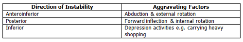
Shoulder instability is a symptomatic abnormal motion of the Glenohumeral Joint, which can present as pain or a sense of displacement (subluxation or dislocation) (Jaggi & Lambert 2010).  96% of shoulder dislocations are attributed to a traumatic event, and 4% due to minor injury or repetitive use.  Following injury patients may develop both structural, and non-structural components of instability, which must be recognised if management is to be successful (Jaggi & Lambert 2010).  The structural instability (usually capsuloligamentous and/or labral pathology) can be thought of as a static instability problem whereas the non-structural indicates a dynamic stability problem, i.e. muscle patterning issues.

In the past shoulder instability has been classified as TUBS (Traumatic Unilateral Bankart Lesion Surgery) and AMBRI (Atraumatic Multidirectional Both shoulders Rehabilitation and Inferior capsual shift surgery).  More recently the Royal National Orthopaedic Hospital, Stanmore has expanded this into three groups. The Stanmore Triangle is a useful classification system which also helps guide management.

The Stanmore Triangle

Polar type I
History of significant traumatic dislocation (most often in anterior direction), usually requiring relocation in A&E. In general they present with; positive apprehension test, and weakness in rotator cuff, especially subscapularis. Global pressure, single leg stance and scapular control are often undisturbed (Jaggi & Lambert 2010).  There will be structural pathology.

Polar type II
Harder to define. In general they present with positive apprehension, increased capsular laxity (excessive external rotation an sulcus sign) and often Glenohumeral internal rotation deficit (GIRD). Global posture may initially appear ok but if further challenged, ie single leg stance, four point kneeling problems with trunk and scapular stability may become more apparent.
There will be a structural lesion (though less severe than type I). May or may not have history of minor trauma, as structural elements may be congenitally abnormal or acquired microtraumatic lesions over time. Patients may describe possible subluxation(s) rather than true dislocation.

Polar type III
No distinct trauma. Dislocation/sublaxation can be voluntary (party tricking) or involuntary. There is no structural defect, instability is due to muscle patterning, with over activity in large muscle groups; lats dorsi, pec major and anterior deltoid and under activity/suppresions of rotator cuff. It is usually muscle recruitment/ timing problem rather than a true weakness.
There is often laxity to the Glenohumeral Joint in all directions and generalised hypermobility. Global posture, trunk and scapular stability are often disrupted.
Classification may not be distinct Polar type I, II, and III, but may be a combination of more than one pole. There is also the ability to move between poles. In addition it is also important to establish the direct/directions of instability.
Dislocation table

Clinical testing

Apprehension (Video link 23s) (link correct as of 12/07/19)

The patient is positioned in supine, with the shoulder abducted to 90° and the elbow flexed to 90°. The clinician stands alongside the patient, supporting the upper limb and externally rotates the shoulder towards end of range.  A positive result is indicated if the patient reports a feeling of instability, apprehension, vulnerability or pain during movement, as the external rotation force is applied.
In the case of a positive test then proceed to the relocation test.

Apprehension and Relocation (Fowler’s Test) (Video link 29s(link correct as of 12/07/19)

The procedure of the test follows the same principles and positioning as for the apprehension test, however when the patient is at the point of apprehension a the clinician applies a posteriorly directed force to the humeral head and asks the patient if this reduces the feeling of apprehension. A positive result is indicated by the patient reporting a reduction of the feeling of apprehension.


Anterior drawer (Video link 45s) (link correct as of 12/07/19)

This test was proposed as useful in patients with a painful shoulder where the apprehension test is difficult to interpret. Ideally this test should be performed with the patient in supine. The examiner stands facing the affected shoulder e.g. left. They fix the patient's left hand in their right axilla by adducting their humerus. The affected shoulder  is held at 80-120° of abduction, 0-20° of forward Flexion and 0-30° of external rotation. The examiner holds the patients scapula spine forward with his index and middle fingers; the thumb exerts counter pressure on the coracoid. The scapula is fixed. The examiner uses his right hand to grasp the patient's relaxed upper arm and draws it (NB it is possible to repeat the anterior drawer in different positions of abduction and external rotation). A positive result is where the relative movement between the fixed scapula and the moveable humberus can easily be appreciated and graded. Occasionally the examiner may reproduce an audible click on forward movement of the humeral head due to labral pathology, and this is ususally associated with apprehension.


Posterior Drawer (Video link 34s) (link correct as of 12/07/19)

Patient positioned in supine with the shoulder positioned in 100-120° abduction, and slight flexion. The elbow positioned in 120° flexion. The clinician stands alongside the patient, with one hand supporting the patients scapula by grasping over the top of the shoulder. The other hand takes a firm hold of the elbow. The clinician then applies a medial rotation movement and a posteriorly directed force to the shoulder joint, transmitted along the length of the humerous. The clinician can then vary the degree of abduction, flexion and medial rotation to try and identify the position of instability. A positive result is indicated by an increased degree of posterior translation of the humeral head on the glenoid of the scapula.


Sulcus
Patient in sitting with their arm relaxed by their side. The clinician grasps the arm at the elbow with one hand and stabilises at the scapula with the other. A gradual inferior pull, is applied to the humerous, attempting to pull the humeral head down on the glenoid fossa. A positive result is indicated by identification of a subacromial sulcus (abnormal dip under the acromion on the lateral aspect of the shoulder). This may suggest inferior instability of the Glenohumeral Joint.

Rotator Cuff integrity testing
As the shoulder dislocates there is the potential for damage to the rotator cuff, therefor cuff integrity should be checked as early as possible. Refer to shoulder impingement syndrome testing for details of rotator cuff tests, (and management should these tests be positive).


GIRD (Glenohumeral Internal Rotation Deficiency, posterior capsule).
Refer to shoulder impingement guidelines.

Imaging

Where there has been a traumatic injury X-rays are frequently carried out in A&E, to confirm the diagnosis, rule out associated fractures and confirm Glenohumeral alignment post relocation.

Magnetic resonance arthrography (MRA) and/or diagnostic arthroscopy can be useful in those suspected to have a structural problem, (Polar type I and II) (Jaggi & Lambert 2010).

Management

Overview

  • Optimise pain management
  • Rehabilitation as appropriate

Patient centred care

Treatment should take into account individual patient needs, preferences, expectations and functional status.  Clinical reasoning should inform treatment based on subjective and objective findings. Good communication between therapist and patient is essential if a successful outcome is to be achieved. Treatment options should be clearly explained so that patients can make informed decisions with regard to their care and management.

Local consultants protocol / guidance

For patients referred by A&E/Orthopaedics and remaining under their care, local consultant protocols/guidance should be followed. This includes those referred with fracture/disclocations.
The Stanmore Triangle allows physiotherapist to reason out a management strategy.

Type I, acute primary dislocator

No evidence to support the use of slings, however can be used for comfort for the first week if required.

Analgesia should be optimised to enable compliance with rehabilitation programme.

An exercise programme should aim to; regain full range of movement and address any issues of scapular stability/control, rotator cuff strength/control and neuromuscular control (as found on assessment). There is rarely an issue with muscle patterning however if found this should be addressed. 

Later stage rehabilitation should include sport/job specific exercise/activity.

Type II and III

Analgesia should be optimised to enable compliance with rehabilitation programme.

An exercise programme should aim to; regain full range of movement and address any issues of scapular stability/control, rotator cuff strength/control and neuromuscular control. There is often (Type II)  / always (Type III) an issue with muscle patterning and if found this should be addressed, by facilitating core stability, anterior/posterior slings and rotator cuff as deemed appropriate alongside muscle lengthening.

Progression and escalation

Progressing as expected (up to 3 Rxs) before discharge or onward referral. 

Refer to ortho, GP, other

Early Escalation (in conjunction with continued MSK Physiotherapy)
Due to the high risk of re-dislocation, traumatic (Polar Type I) young (under 27), athletic and/or manual worker should be referred early to orthopaedics. As the majority of this group tend to attend A&E they are usually referred on at this time, however a referral has not been made or the patient comes directly to physiotherapy an early referral to orthopaedics is indicated.
Over 40 – 75 with evidence of rotator cuff tear and patient wishes to consider surgical intervention early refer to orthopaedics (refer to rotator cuff pathway).

Escalation outwith MSK Physiotherapy
At any point during the rehabilitation process if the patient and physiotherapist assess that progression is hindered by pain, poor control of the shoulder, continued instability/subluxing or further dislocations, referral to orthopaedics should be considered.

Recurrent dislocator, with ongoing apprehension or instability and no evidence of muscle patterning, (abnormal muscle patterning can account for 45% of recurrent shoulder instability (Malone et al 2006)).

Referral to orthopaedics should only be considered if the patient wishes to consider surgical options.

Evidence

Day R, Fox J and Paul-Taylor G. 2009. Neuro-Musculoskeletal Clinical Tests A Clinicians guide, Churchill Livingstone ISBN 9780443069451.

Farber, A.J., Castillo, R., Clough, M., Bahk, M., McFarland, E.G. 2006. Clinical Assessment of 3 Common tests for traumatic Anterior Shoulder Instability.  Journal of Bone and Joint Surgery 88 (7): 1467-1480. Link Here (link correct as of 18/03/2024)

Hayes, K. et al. (2002) Shoulder Instability: Management and rehabilitation, Journal of Orthopaedic Sports Physical Therapy 32(10); 497-509.

Jaggi, A., Lambert, S. (2010) Rehabilitation for shoulder instability.  British Journal of Sports Medicine 44(5): 333-340. Link Here (link correct as of 18/03/2024) 

Lewis, A., Kitamura, T., and Bayley J.I.L. 2004. The classification of shoulder instability - new light through old windows.  Current Orthopaedics 18: 97-108. Link Here (link correct as of 18/03/2024) 

Malone, A.A., Jaggi, A., Calvert, P.T., Lambert, S., Bayley, I. Muscle Patterning Instability: Classification and Prevalence in a Referral Shoulder Service. In: Norris, T.R., Zuckerman, J.D., Warner, J.J.P, Lee, T.Q., editors .2006. Surgery of the Shoulder and Elbow : An International Perspective. USA: American Academy of Orthopaedic Surgeons.

Stanmore Classification for Shoulder Instability by Ian Bayley January 2014 http://www.youtube.com/watch?v=2Hl3sP1-oVs (link correct as of 18/03/2024).

Patient information

NHS GGC Physiotherapy Service - shoulder https://www.nhsggc.scot/hospitals-services/services-a-to-z/musculoskeletal-msk-physiotherapy/msk-self-help-and-information-shoulder/

NHS Inform may provide useful information, especially for those presenting to physiotherapy within the time frames of natural recovery.
https://www.nhsinform.scot/illnesses-and-conditions/muscle-bone-and-joints/self-management-advice/shoulder-problems (link correct as of 18/03/2024).

Patient education in terms of times frames and prognosis is paramount. Consider use of local/national publications to support.

Editorial Information

Last reviewed: 30/04/2024

Next review date: 30/04/2025

Reviewer name(s): Louise Ross, Alison Baird, Karen Glass.