Giant Cell Arteritis (GCA) (Guidelines)

Warning

Initial referral is either to ambulatory emergency care or if there is visual loss or amaurosis fugax to ophthalmology. Ultrasound/ biopsy (if available) and initial treatment will be coordinated by the teams above. A follow up appointment will be generated in rheumatology. CRP/PV should be taken before steroids started.

Presentation

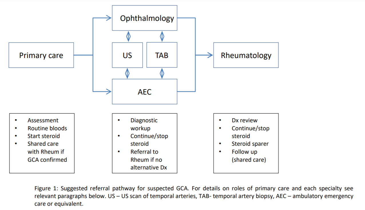
GCA should be suspected:

  • In individuals over 50 years old (usually over 60 years old) with new onset headache
  • And other typical symptoms and signs of GCA (see list below)
  • And elevated inflammatory markers
  • And no alternative explanation for the patient’s presentation.

It is very rare for GCA to present with normal inflammatory markers and we expect either CRP, plasma viscosity (PV) or both to be raised. (There is no data on performance of plasma viscosity which has replaced ESR in NHS Highlands, but studies suggest less than 3% GCA cases have normal CRP and ESR).

There is no validated clinical scoring system to estimate the likelihood of GCA. In general, the more symptoms/signs present and in the absence of an alternative diagnosis, the greater the probability of GCA. None of the symptoms or signs is specific or pathognomic for GCA, and its mimics include many other local, systemic, infectious and malignant conditions. Patients with suspected GCA will therefore require further work-up to confirm a diagnosis.

GCA is a medical emergency requiring urgent specialist assessment but investigations should not delay initiation of steroid treatment if GCA is strongly suspected.

Symptoms and signs

Symptom / Sign Notes

New onset of unaccustomed, persistent, localised headache: 

Typically unilateral, most commonly temporal, but can be more widespread or affect occipital area only.

Constitutional symptoms:

Eg. fatigue, night sweats, anorexia, weight loss, fever, depression - present in most cases.

Scalp tenderness:

In about 50%, eg. sensitivity to touch, eg. when brushing or combing hair.

Tenderness, thickening or nodularity of temporal artery: 

(Present in 45 to 75%) with or without decreased pulsatility.

Jaw claudication: 

Pain on chewing or difficulty chewing. Potentially very specific but to be differentiated from TMJ disorders and other mimics eg. myasthenia gravis and parotid tumours. Tongue pain and claudication – rare, causes difficulty talking.

Sudden, profound, painless visual loss: 

Initially unilateral, but may rapidly become bilateral. May be preceded by transient visual obscurations or longer amaurosis fugax-like episodes. Sudden onset of significant double vision (rare).

Polymyalgia rheumatica (PMR) symptoms: 

Up to 40% of cases will have PMR symptoms: shoulder and pelvic girdle stiffness, early morning stiffness, diurnal pattern.

Neurological features (rare): 

Mono or poly neuropathy of limbs, cranial nerve palsies, carotid of vertebrobasilar stroke/TIA.

Audiovestibular symptoms, dry cough, sore throat, hoarseness (very rare).


Extra-cranial ischaemic features (rare):

Eg. limb claudication with reduced peripheral pulses or asymmetric arm BP, bruits over subclavian or axillary arteries, carotidynia.

Differential Diagnosis

Consider:

  • Headaches (tension, cluster, migraine, trigeminal neuralgia, etc).
  • Acute infections eg. Herpes zoster, meningitis.
  • Acute angle closure glaucoma.
  • TIAs and embolic visual deficits.
  • TMJ and dental pain, sinus disease, ear problems.
  • Cervical spondylosis and other upper cervical spine disease.
  • Myeloma with cervical or cranial deposits, or other cranial malignancy.
  • Serious intracranial pathology, such as haemorrhage, retro-orbital or base of skull lesions.
  • Other types of vasculitis.

Referral Pathway

Role of the primary care

  • Urgent clinical assessment of patient presenting with suspected GCA.
  • Document history, symptoms and signs.
  • Routine bloods prior to starting steroid - minimum set: FBC, U+Es, LFTs, CRP, plasma viscosity (ESR if available), glucose/HbA1c if patient not known to be diabetic.
  • Immediate referral to/discussion with Raigmore AEC or local acute medical team for further diagnostic workup* .
  • Start steroid treatment (see treatment section) if GCA strongly suspected. It may be appropriate to start before blood tests available. Document start date and start dose in a referral.
  • Further follow-up in accordance with shared care model (see Follow-up, Treatment and Relapse sections for details).

*In case of acute significant visual symptoms- as outlined above- refer urgently to ophthalmology for assessment and consideration of pulsed IV methylprednisolone.

Role of the Acute Medical Team

  • Clinical assessment with differential diagnosis in mind (see above; also consider checking for other large vessel involvement: subclavian/axillary bruits, difference in BP both arms, asymmetry in peripheral pulses).
  • Diagnostic work-up to help confirm or exclude GCA/other pathology (e.g. CT head and/or LP, CXR, urinalysis, calcium, immunoglobulins/Bence-Jones protein, if PV/ESR disproportionate to CRP).
  • If GCA likely and no safe alternative diagnosis identified: refer for US of temporal/axillary arteries to US department (please use radiology referral form). If there is no availability contact rheumatology secretaries: (01463 706556 or 705443 or email louise.macleod2@nhs.scot or julie.davidson.3@nhs.scot); referral form for US of temporal arteries.
    US should be organised within 7 days from starting steroid and ideally within 24 to 48 hours. Please note that due to insufficient resources US scanning capacity may be limited.
  • When US not available within 7 days from starting steroid or result non-conclusive: refer for TAB to vascular surgeon in Raigmore or general surgery in other hospitals depending on patient’s location. The patient must be counselled and given the patient information leaflet 'For patients referred to TAB'. Referral form for temporal artery biopsy to be passed to Mr Bernhard Wolf (Raigmore cases)
    Please note that sensitivity of TAB in patients started on high dose steroid declines with time; biopsy should be done within 2 weeks from starting steroid.
  • Make provisional diagnosis and decision re initiation/continuation of steroid based on clinical assessment, results of investigations +/- response to therapy
  • If there is diagnostic uncertainty or if the patient may require steroid sparing therapy please discuss with duty rheumatologistvia rheumatology secretaries: (01463 706556 or 705443 or email louise.macleod2@nhs.scot or julie.davidson.3@nhs.scot)
    Note that performance of US and TAB in GCA diagnosis is variable and largely depend on clinical pre-test probability and timing of the test. Therefore decision making must be based upon clinical assessment, results of investigations, absence of alternative diagnoses and the response to therapy, which is typically rapid but can be up to 2 weeks. As a general rule positive TAB confirms the diagnosis but negative TAB does not necessarily exclude it. Sensitivity of TAB varies between studies from 40 to 80%. In general, accuracy of US supports its use either for ruling out GCA in low-probability cases or for confirming GCA in high-probability cases but in all other scenarios additional efforts towards a diagnosis are necessary. Patients with jaw claudication and CRP greater than 40 mg/L have a 93% probability of having GCA. See section: clinical decision aid.
  • Direct patient to https://www.versusarthritis.org/ (or hand out printed leaflet) for information about GCA diagnosis and its treatment. The patients referred for TAB should be given the patient information leaflet  'For patients referred to TAB'.
  • Agree treatment plan with GP and steroid taper regimen (see treatment section).
  • Urgent referral to Rheumatology for review (see follow-up section).

Treatment

  • Start 40 to 60 mg prednisolone immediately if strong suspicion of GCA and continue once daily.
    • Initial dosing is not evidence based. Lower doses might be appropriate in smaller patients or those at greater risk of side effects from systemic steroids eg. diabetes, severe osteoporosis and especially in the absence of ischaemic symptoms (jaw/tongue claudication, TIA/stroke, visual loss); higher doses might be considered in larger patients or those with ischaemic symptoms;
    • Patients with visual symptoms or loss should be referred urgently (same day) to ophthalmology and may require initial pulsed IV methylprednisolone. In hours, this treatment is the responsibility of ophthalmology. Out of hours, AEC or acute medical team to organise first 0.25 to 1 g IV methylprednisolone pulse and discuss further management with ophthalmologist next day.
  • Once GCA controlled (symptoms resolved, normal inflammatory markers) start tapering steroid to target dose 15 to 20 mg/d within 2 to 3 months and 5mg or less within 1 year. Recommended duration of treatment with steroid alone is 1 to 2 years, providing no relapse (EULAR); discontinuation of steroid monotherapy in under 1 year is associated with higher risk of relapse;
    • There is no evidence-based tapering regimen; initial reduction could be for example by 10 mg every 2 weeks down to 20 mg, then by 2.5 to 5 mg every 2 weeks down to 10 mg/d, then by 1 mg every 1 to 2 months. Clinical response with normalisation of inflammatory markers should happen within 7 to 14 days in majority of patients; if there is no symptom resolution/normalisation of inflammatory markers within this timeframe then re-evaluation of diagnosis is necessary.
  • GP to assess and manage co-morbidities relevant to treatment, such as diabetes mellitus, hypertension, and risks of osteoporosis, infection and cardiovascular disease.
    • All patients to be started on oral bisphosphonate + 800 units Vitamin D +/- calcium, if not contraindicated.
    • DEXA in patients under 65 years.
    • Routine use of aspirin or statin not advised unless indicated for other reason.
    • Decision regarding gastroprotection with PPI inhibitor should be based on individual risk.
    • For those at risk of developing secondary diabetes/hyperglycaemia, or in those with pre-existing diet or tablet controlled diabetes who are not glucose monitoring, consider referring to practice nurse or Diabetes Specialist Nurse to start glucose monitoring; initially monitoring glucose levels once daily at approximately 5pm.
    • Patients with ocular hypertension/at risk of glaucoma should be assessed by optometrist or ophthalmologist.
    • Live vaccinations are contraindicated in patients on more than 20mg/d prednisolone and on DMARDs (except Zoster vaccine in patients on moderate dose of methotrexate).
    • Patients without a history of chicken pox (varicella zoster virus infection) should be advised to avoid close contact with people who have chickenpox or shingles, and to seek urgent medical advice if they have been exposed.
  • In patients at high risk of steroid toxicity (eg. poorly controlled diabetes, severe osteoporosis, psychosis) or who relapse, adjunctive therapy with tocilizumab or methotrexate can be considered: please discuss with rheumatology. In patients on steroid-sparing agents tapering protocol might be faster - see: steroid tapering protocol for patients on tocilizumab .
Steroid tapering protocol for patients on tocilizumab
WeekDose mg/dWeek Dose mg/dWeek Dose mg/d
1601012.5194
2501110204
340129213
435138223
530147232
625156242
720166251
815175261
912.5185270

Follow-up

  • Urgent rheumatology appointment within 4 to 6 weeks for review of diagnosis and requirement for steroid sparing therapy; referrals will be taken from acute medical team and ophthalmology,
  • Primary Care follow-up as per shared-care model:
    • 2 weeks and 4 weeks then
    • every 2 months for first six months then
    • every 3 months thereafter in first year then
    • every 3 to 6 months in second year, and additionally as required (eg. in case of relapse).
  • GP will be responsible for:
    • Routine bloods monitoring prior to steroid dose reduction, in particular CRP/PV.
    • Monitoring for evidence of relapse and disease-related complications (see below)
    • Attention to any new features that might suggest alternative diagnosis.
    • Corticosteroid-related complications eg. osteoporotic risk and fractures, diabetes.
    • For those at risk of developing secondary diabetes/hyperglycaemia, or in those with pre-existing diet or tablet controlled diabetes who are not glucose monitoring, consider referring to practice nurse or Diabetes Specialist Nurse to start glucose monitoring; initially monitoring glucose levels once daily at approximately 5pm.
    • Where applicable, methotrexate monitoring.
  • Rheumatology review at 3 to 6 month intervals in selected patients
    • With relapsing disease or on a steroid-sparer
    • When required eg. in case of suspected relapse. Rheumatology will be responsible for organising tocilizumab monitoring where ITR facilities exist.

Relapse

Relapse is defined as return of GCA symptoms+/- signs with a rise in inflammatory markers.

  • Minorrelapse: typical cranial symptoms without ischaemic features:
    • Increase prednisolone back to previous dose that controlled the symptoms or by 5 to 15 mg, and restart tapering once disease is controlled again.
  • Majorrelapse: ischaemic features present eg. jaw or tongue claudication, visual loss, TIA/stroke:
    • Increase prednisolone back to 40 to 60mg
    • In case of acute visual loss or significant diplopia refer urgently to ophthalmology for possible IV pulse methylprednisolone.
    • Restart tapering once disease is controlled again.
  • In all cases of suspected relapse:
    • GP to contact acute medical team to discuss need for further imaging/TAB and adjustment of therapy; acute medical team will refer for US/TAB if required and inform rheumatology who will see urgently in clinic within 4 weeks
  • Inability to reduce steroid, persistent constitutional symptoms, PMR symptoms or persistent elevation of inflammatory markers may indicate another diagnosis or involvement of aorta and other extra-cranial vessels – discuss with rheumatology.
  • A rise in inflammatory markers should increase suspicion of relapse but does not confirm it.
  • Arthralgia/myalgia/lethargy without rise in inflammatory markers can be due to steroid withdrawal and should not lead to an increase in steroid dose unless there are other symptoms/signs suggestive of relapse – if concern discuss with rheumatology.

Clinical decision aid

Possible approach to clinical decision making for GCA diagnosis based on clinical probability and results of US/TAB as per BSR recommendations 2019

Probability of GCA is based on clinical judgement using all information available (symptoms, signs,  laboratory tests, and alternative non-GCA explanations for the clinical picture) and can be updated based on new information (clinical course, result of temporal and axillary ultrasound and/or result of temporal artery biopsy). This assessment should ideally be performed by an individual with specialist expertise. Note that for a medium estimated probability of GCA, it may be useful to perform an ultrasound prior to biopsy, in case the biopsy is negative

Abbreviations

Abbreviation Meaning
AEC Absolute eosinophil count
CRP C-reactive protein
CXR Chest x-ray
ESR Erythrocyte sedimentation rate
FBC
Full blood count
GCA Giant Cell Arteritis
IV Intravenous
LFT Liver function test
PMR Polymyalgia Rheumatica
PV Plasma viscosity
TAB Temporal artery biopsy
TIA Transient ischaemic attack
TMJ Temporomandibular joint
U+E Urea and electrolytes
US Ultrasound

Editorial Information

Last reviewed: 20/01/2023

Next review date: 31/01/2026

Author(s): Rheumatology Department .

Version: 8

Reviewer name(s): Consultant Rheumatologist.

Document Id: TAM481