Warning

Background

Abdominal quadrant regions
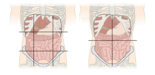
Abdominal quadrant regions by Jmarchn is licensed under CC BY-SA 3.0

Blunt abdominal trauma is more challenging than penetrating trauma, where the decision to operate is usually straightforward. All abdominal trauma is managed one of three ways:

  1. Operative
  2. Non-operative management (NOM) with IR
  3. NOM without IR

Pitfalls!

  • Suspect rather than rely on numbers - abdominal haemorrhage is often concealed, is always non compressible and recognition is challenging.
  • The surgical hand will only detect 50% of abdominal injuries, even in conscious patients
  • Beware – elderly, obese, obtunded and spinal cord injuries
  • This starts at the nipples anteriorly and scapular angle posteriorly, though trauma rarely respects these boundaries.
  • Do not remove the pelvic binder to perform a laparotomy
  • Patients with a sustained SBP <70mmHg and abdominal trauma should probably be in theatre.
  • Ureteral injuries are rare but commonly missed

Surgery

  • A midline laparotomy should be used over other approaches
  • Haemorrhage control should occur as quickly as possible within 1 hr of presentation.
  • A surgical consultant should be present for all laparotomies in trauma.
  • The following are the commonest indications for an immediate laparotomy.
    • Unstable patient with positive FAST
    • Peritonitis
    • Unstable patient with free fluid on CT
    • Hollow viscus injury
    • Retained weapon
    • Gunshot wound
    • Evisceration
  • A laparotomy should be strongly considered when free fluid is present in the absence of solid visceral injury

Damage control

  • Utilise damage control principles (proximal control, haemostasis and faecal/urinary diversion) over definitive procedures in selected patients with physiological compromise.
  • Consider temporary abdominal closure in the presence of physiological compromise.
  • The second look should follow between 24-72 hours after the first operation.

CT
CT is the gold standard imaging modality of choice in blunt abdominal trauma

FAST

    • Unstable patients
    • Multiple casualties where triage can be challenging. A FAST should can be useful in the following situations
  • FAST can rule in intra-abdominal haemorrhage
  • A negative FAST does not rule out injury
  • A negative FAST does not rule out haemorrhage!

    Ultrasound
    Ultrasound should not be used acutely to assess intra-abdominal injury in adults. CT is modality of choice.

Penetrating abdominal trauma specifics

Imaging

  • Perform CT in all penetrating trauma where Non-operative Management (NOM) is being considered.
  • NOM should be considered if there is all three of:
    • Haemodynamic stability
    • Absence of peritonitis
    • Absence of diffuse abdominal tenderness (away from wound)

Laparoscopy

  • Consider in left sided thoracoabdominal injuries to rule out diaphragmatic injury
  • Can be used to determine peritoneal penetration

Antibiotics

  • Administer a single pre op dose of antibiotics as per local guidelines for all patients undergoing a laparotomy for penetrating trauma.
  • If there is no hollow viscus injury then no further antibiotics are required
  • If there is hollow viscus injury then continue antibiotics for 24 hours
  • Consider repeat dosing of antibiotics during massive transfusion.

Penetrating colon injuries

  • Resect penetrating colonic injuries and strongly consider leaving definitive repair or stoma formation until relook laparotomy in damage control setting.

Penetrating rectal Injuries

  • Perform proximal diversion in patients with suspected non-destructive penetrating extra-peritoneal rectal injuries
  • Do not use presacral drains or perform distal rectal washout.

Discharge of penetrating injuries
Discharge after 24 hours in the presence of reliable clinical examination and minimal to no abdominal tenderness

Penetrating colon injuries
Resect penetrating colon injuries and consider colostomy during damage control.

Penetrating rectal injuries

  • Perform proximal diversion in patients with non-destructive penetrating extra-peritoneal rectal injuries
  • Do not use presacral drains or perform distal rectal washout

Discharge of penetrating injuries
Discharge after 24 hours in the presence of reliable clinical examination and minimal to no abdominal tenderness

Algorithm for abdominal trauma

Algorithm for penetrating trauma


Splenic trauma

Operative management – splenectomy indications:

  1. Separate indication for laparotomy
  2. Unresponsive haemodynamic instability
  3. Ongoing signs of haemorrhage after IR 

Non-operative management (NOM) indications:

Any grade of injury even with free fluid and pseudoaneurysm.

Only consider in patients with severe traumatic brain injury when there is immediate access to IR and surgery.

Risk factors of NOM failure:

  1. Age >55yrs
  2. Grade III, IV & V injuries
  3. High injury severity score
  4. Large hemoperitoneum
  5. Hypotension before resuscitation
  6. GCS<12
  7. Low haematocrit on admission
  8. Blush on CT
  9. Anticoagulated
  10. HIV
  11. Drug addiction
  12. Cirrhosis
  13. Blood transfusion required

Interventional radiology

Absolute indications

  • IR is the 1st line intervention where there is active arterial extravasation, regardless of injury grade and stability and no other indication of laparotomy.

Strong consideration in:

  • Stable/stabilising grade III-V injuries with active arterial extravasation, pseudo-aneurysm or AV fistula.
  • All grade IV/V injuries
  • Persistent signs of haemorrhage and isolated splenic injury on CT, regardless of the presence of active arterial extravasation.
  • Grade III injuries in the presence of risk factors for NOM failure
  • Blush seen on CT but not present during angiography

Admission and ongoing management

  • Transfer patients with a grade III/IV/V to the MTC for a minimum of 48-72 hours observation and bed rest.
  • Start LMWH 24 hours after injury in most cases, even in patients undergoing NOM
  • Reversal of anticoagulant should be individualised on risk-benefit
  • Consider repeat CT scanning during admission in:
    • Grade III-V injuries
    • Decreasing haematocrit
    • Presence of blush, pseudoaneurysm or AV fistula on initial scan
    • Underlying splenic pathology*
    • Coagulopathy*
    • Neurologically impaired patients

Vaccinations 

  1. See guidance in the Splenectomy and Dysfunctional Spleen Prophylaxis for Adults and Children guideline.
  2. Give pneumococcal, Hib, Men C and influenza vaccines after 14 days or upon discharge

In embolised patients immune function is thought to be preserved and no antibiotic or vaccine prohpylaxis is required

Liver trauma

Surgical principles

Absolute indications

  • Haemodynamic instability
  • Peritonitis
  • Penetrating injury with any of:
    • significant free air
    • localised thickened bowel wall
    • Evisceration
    • Impalement
    • Free fluid without solid visceral injury
    • Other organ injury requiring laparotomy 

Relative indications

Severe head or spinal cord injury which impairs clinical assessment

Non-operative management (NOM)

  • Should be attempted in all grades of liver injury not requiring laparotomy
  • All grade III/IV/V injuries and all penetrating liver trauma should be monitored in the MTC:
    • 4-6hrly bloods and clinical examination for 48hrs in a monitored environment 

IR

Indications (In the absence of another indication for laparotomy)

  • Stable patients with active arterial extravasation on CT
  • Unstable patients with active arterial extravasation responding to resuscitation
  • Hepatic artery pseudo aneurysms
  •  Post operative repair for liver injury 

IR can be safely repeated if required.

Complications of liver trauma
12-14% complication rate, most commonly in grade IV/V consisting of:

  • Bleeding
  • Abdominal compartment syndrome
  •  Abscesses
  • Necrosis
  • Biliary complications:
    • Leak
    • Haemobilia (raised bilirubin +/- upper GI bleed)
    • Biloma
    • Peritonitis
    • Fistula 

Further imaging / interventions

Perform a repeat CT in the presence of

  • Abnormal inflammatory response
  • Abdominal pain
  •  Fever
  • Jaundice
  • Drop in Hb
    • Biliary complications may require ERCP, stenting, drainage or surgery

Kidney trauma

History & examination

  • Pre-existing renal pathology/surgery makes injury more likely
  • The following examination findings may suggest renal injury:
    • Flank pain
    • Fractured lower ribs
    • Abdominal tenderness/distension/mass

Investigations

Perform a Urinalysis and Creatinine in all patients.

Imaging:

USS
USS is not useful in the acute setting

IVP

  • IVP should only be used if CT is unavailable
  • Perform a one shot IVP in the OR for patients who were taken directly to theatre without imaging (2ml/kg of contrast followed by plain film after 10 mins.)

 CT

  • CT is the imaging modality of choice and indicated in the presence of:
    • Frank haematuria
    • Microscopic haematuria with a single episode of hypotension
    • All penetrating Trauma
  • Consider in the following, even in the absence of haematuria:
    • Rapid deceleration injury
    • Direct flank trauma
    • Flank contusions
    • Lower rib/thoracolumbar fractures

Concerns regarding contrast worsening outcomes are unwarranted as low rates of contrast induced nephropathy are seen in renal trauma patients

A delayed phase scan should be performed in all renal injuries to identify urine leak 

Non-operative management (NOM)

  • All injuries can be attempted to undergo NOM.
  • Pedical and vascular avulsion injuries often require surgery +/- IR
  • Transfer all patients with grade III/IV/V injuries to the MTC for serial 6 hourly clinical and laboratory observation for 24 hrs in a monitored environment.

Interventional radiology

  • All injuries can be attempted to undergo NOM.
  • Pedical and vascular avulsion injuries often require surgery +/- IR

Transfer all patients with grade III/IV/V injuries to the MTC for serial 6 hourly clinical and laboratory observation for 24 hrs in a monitored environment.

Indications

  • Active extravasation of contrast at WBCT / CT angiography.
  • AV fistula
  • Pseudonaeurysm
  • Some blunt grade III
  • Grade IV/V injuries
  • Penetrating injuries 

Surgery
Absolute Indications

  • Haemodynamic instability
  • Expanding or pulsatile haematoma seen at laparotomy
  • Patients who have an abnormal one shot IVP during laparotomy

Relative indications

  • Grade V blunt vascular injuries involving renal pedicle or avulsion*
  • Grade IV or V penetrating injury*

*Senior Consultant input with urology/IR if NOM is to be considered

Nephrectomy is the procedure of choice in damage control / major haemorrhage.

Only attempt renal reconstruction if haemorrhage controlled and there is sufficient viable renal parenchyma

Antibiotics
Please refer to the antimicrobial guidelines here

Repeat imaging

  • All grade V injuries after 72 hours
  • At 48-72hrs if urinary extravasation seen on initial scan to determine need for diversion procedure
  • Fever with no other explanation
  • Decreasing haematocrit
  • Significant flank pain 

Ongoing management / follow-up

  • Bed rest until haematuria is light and bladder irrigation not required
  • Follow up after 3 months, monitoring blood pressure, creatinine and urinalysis.

Pancreatic injuries

Investigations / imaging

  • CT scan is the diagnostic modality of choice
  • Raised amylase is suggestive of but not diagnostic of pancreatic injury

Principles

  • Transfer all pancreatic injuries to the RIE
  • Strategies include:
    • Operative
    • NOM
    • Drainage
    • Suture repair
    •  Resection for major injuries.
    • Endoscopic stenting
  • In damage control situations perform drainage only

Management

Grade I/II injuries (no ductal involvement seen on CT)

  • 1st line is NOM
  • Evaluate further with MRCP/ERCP as this may change the grade/management
  • If diagnosed at laparotomy use non-resectional management:
    • pancreatography, drainage or no drainage 

Grade III/IV (any ductal involvement)

  • Operative management has fewer complications than NOM and is recommended.
  • If diagnosed at laparotomy undertake resection (drainage if damage control) 

Octreotide is not recommended as postoperative prophylaxis to prevent fistula.

Antimicrobial Guidelines

Please refer to the antimicrobial guidelines here.

Editorial Information

Last reviewed: 01/09/2021

Next review date: 01/09/2024

Version: 1.0