Estimated Due Date (EDD) Ultrasound Scan (501)

Warning
Please report any inaccuracies or issues with this guideline using our online form

EDD should be calculated from the first scan ≥ 11+ 2 weeks gestational age (≥ 45mm).

If the initial scan is performed < 11+2 weeks gestation, the patient should have a return appointment at approximately 12-13 weeks gestation to perform optimal dating +/- NT measurements.
(Refer to Screening / Non-screening Pathways)

Upper and lower limbs must be assessed as well as cranial anatomy

Parameters to be Used for Calculation of EDD and Associated Downs Syndrome Screening Method

As adapted from Chudleigh, Loughna & Evans (2011) from BMUS.

Parameter 

Parameter used for establishing EDD

Down’s Syndrome Screening method

CRL < 45.0mm

Unable to date. Re-scan at 12-13 weeks gestation

N/A

CRL 45.0mm – 84.0mm

CRL

First Trimester CUBS 

CRL >84mm  and HC< 101.0mm

Unable to date.  Re-scan ≥ 14+2 weeks gestation

No screening until EDD confirmed

HC ≥ 101.0mm – 172.0mm

HC

Second Trimester Bloods only (AFP)

1st Trimester dating (CRL 45.0mm-84.0mm)

Recommended criteria for measurement of CRL for pregnancy dating (UKNSC, 2015):

CRL DETAIL TO BE DEMONSTRATED
MIDLINE SECTION
  • Sagittal section of the fetus with the head in line with the full length of the body
  • Echogenic tip of the nose
  • Rectangular shape of the palate
  • Translucent Diencephalon
  • CRL axis should be between 0-30 degrees to horizontal
  • Clearly defined crown and rump 
POSITION
  • Pocket of fluid, at least equivalent in size to the width of the palate, should be visible between the fetal chin and chest 
  • Fetal palate angle should be 30º to 60º relative to the horizontal 
  • Nasal tip should be level or above the anterior abdominal wall 
MAGNIFICATION
  • Entire CRL section should fill over 60% of the screen 
CALIPER PLACEMENT
  • Correct calliper placement on outer borders of crown and rump
  • Longest length of the fetus should be measured
IMAGE ARCHIVING
  • The CRL should be measured at least twice and the maximum measurement that meets the criteria should be recorded 
  • The image demonstrating the measured CRL which has been reported should be archived

2nd Trimester dating (HC ≥ 101.0mm – 172.0mm)

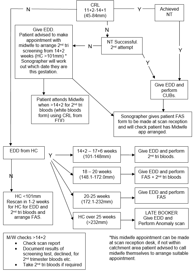
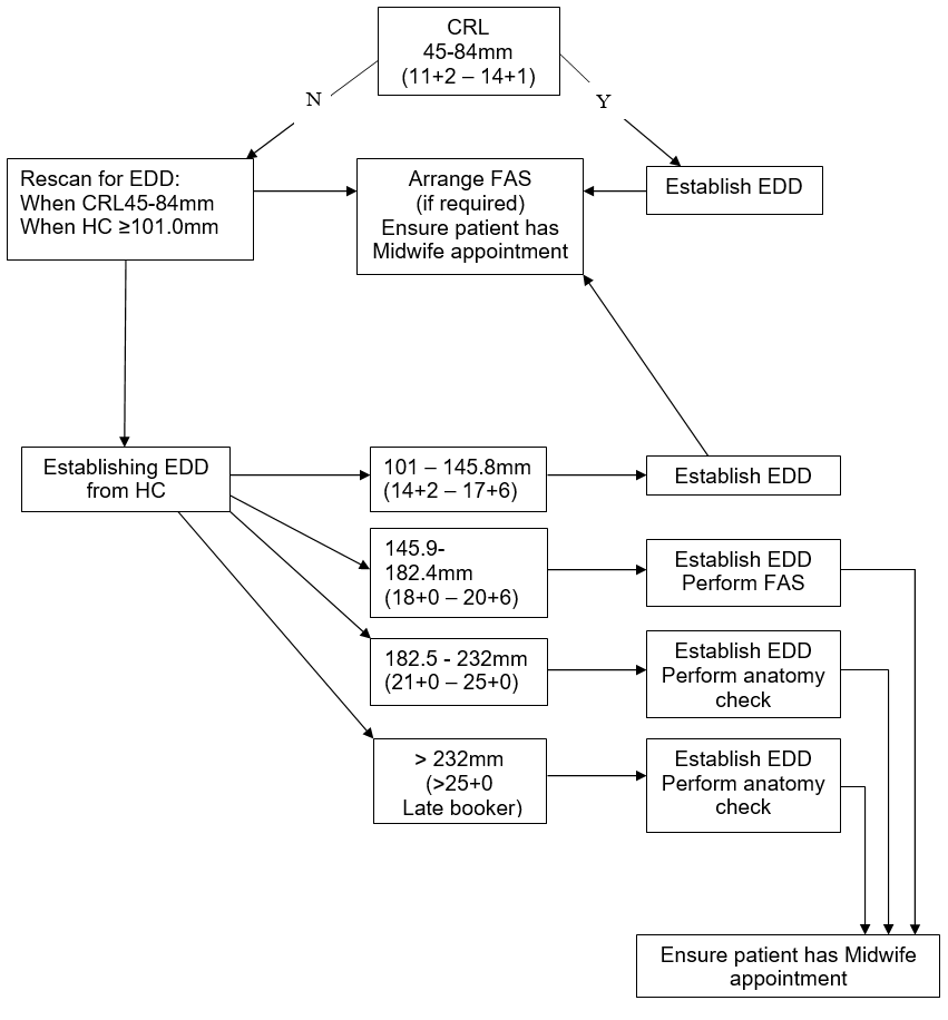
The HC should be calculated using the recommended values of Altman and Chitty as per BMUS recommendations.

Technique for calculation of HC:

A cross-sectional view of the fetal head at the level of the ventricles should be obtained. The image should have the midline echo lying as close as possible to the horizontal plane.

The following landmarks should be identified and the image frozen:

  • rugby ball shape;
  • centrally positioned, continuous midline echo broken at one third of its length by the cavum septum pellucidum;
  • anterior walls of the lateral ventricles centrally placed around the midline;
  • the choroid plexus should be visible within the posterior horn of the ventricle in the distal hemisphere.
  • Callipers should be placed on the outer border of the occipital and frontal bones as close as possible to the midline across the longest part of the skull.

If HC measurements cannot be made then EDD should be calculated using the femur length (FL)

Technique for calculation of FL:

The image should be obtained with the femur lying as close as possible to the horizontal plane. The full length of the bone should be visualised with soft tissue visible at both ends. Calipers should be placed at the centre of the ‘U’-shape at each end of the bone.

Third Trimester (Late Booker: HC > 232mm)

After 25 completed weeks the patient will be classed as ‘LATE BOOKER'.

All three measurement parameters [HC, AC and FL] should be obtained.

An EDD will be calculated from the HC measurement and entered on Badgernet. On the ultrasound report page type in comments ‘EDD based on HC >25 weeks GA’ and create a LATE BOOKER alert.

Multiple Pregnancy

If there is discrepancy in the sizes between the fetuses, the EDD should be calculated from the largest fetus, using the criteria above.

Establishing Gestational Age Prior to Visualisation of a Live Embryo

Gestational age may be assessed from measurements of the mean sac diameter (MSD). This is calculated from the maximal diameters of the gestation sac (in mm) in the longitudinal and transverse views on transvaginal scan. The Mean gestation sac volume should be calculated where a transvaginal examination is declined using the full bladder technique.

An EDD should only be generated once a live embryo or fetus has been identified measuring ≥ 45mm.

Ultrasound EDD/Screening Pathway – Singleton (patients who wish Downs syndrome screening)

Ultrasound: Establishing EDD/Screening pathway - Singleton (Patients who decline T21 screening)

Editorial Information

Last reviewed: 31/05/2022

Next review date: 31/05/2026

Author(s): Donna Maria Bean.

Version: 2

Approved By: Obstetrics Clinical Governance Group

Document Id: 501

References

Altman DG & Chitty LS (1997). New charts for ultrasound dating of pregnancy. Ultrasound Obstet Gynecol 10:174-91

Antenatal Care: Routine care for the pregnant healthy woman. National Institute of Clinical Excellence (NICE) Guildeline.

Chudleigh, T., Loughna, P and Evans, T. (2011). A practical solution to combining dating and screening for down’s syndrome. Education & Training 1:154-71

Fetal Anomaly Screening Programme Handbook for Ultrasound Practitioners. UKNSC  April 2015.

Fetal Size and Dating:Charts Recommended for Clinical Obstetric Practice. BMUS, Jan 2008

Guidelines for Professional Working Standards. Ultrasound Practice. United Kingdom Association of Sonographers. October 2008