Fluid resuscitation of childhood burns

Aim and background

Aim:

To promote a unified approach to fluid resuscitation of children with thermal injuries in Scotland, including the immediate resuscitation in the Emergency Department and further fluid management in specialist units providing ongoing care.

Background:

Thermal injuries affecting more than 20% of the total body surface area (TBSA) in adults and 10% TBSA in children can result in Burn Shock as a result of a combination of electrolyte shifts [Baxter 1968, Moyer 1965, Moylan 1973, Arturson 1979], inflammatory response [Gibran 2000, Scott 2005] and evaporate losses. This sequence of events leads to intravascular hypovolaemia and haemoconcentration that are maximal 12-hours post-injury [Moore 1970]. The clinical sequelae of reduced cardiac output is the combined result of decreased plasma volume, increased afterload, and decreased myocardial contractility. Recent clinical evidence suggests that fluid administration alone is not effective in restoring preload or cardiac output in the first 24 hours post-injury [Holm 2004] which confirms earlier animal-based observations [Baxter 1968]. Under-resuscitation may lead to decreased perfusion, acute renal failure, and death although since the adoption of weight and injury-size based formulas for resuscitation, multiple organ dysfunction caused by inadequate resuscitation has become uncommon [Pham 2008]. Instead, administration of excessive fluid volumes have been reported [Cancio 2004, Engrav 2000, Pruitt 2000] which can lead worsening oedema formation, elevated compartment pressures, Acute Respiratory Distress Syndrome (ARDS), and multiple organ dysfunction [Klein 2007, Sheridan 1994, Sullivan 2006]. In October 2006, the American Burn Association conducted a meeting to determine a research agenda for the next decade. Participants highlighted over-resuscitation as a common, but potentially avoidable phenomenon in today’s burn units [Greenhalgh 2007].

Fluid regimes in the UK

In the UK, burns fluid resuscitation practice has undergone considerable change over the last decade. In 1997, a questionnaire study presented at the European Burns Association annual conference demonstrated that the majority of centres utilised the albumin-based Muir & Barclay regime [Webb 2002] contrary to guidelines promoted during ATLS and EMSB courses that favoured the crystalloid-based Parkland Formula. The following year saw the publication of the Cochrane Injuries Group report that questioned the appropriateness of using albumin in critically ill patients, particularly those with burn injuries [Cochrane Injuries Group Albumin Reviewers 1998]. In 2001, a similar questionnaire demonstrated that over 50% of centres managing paediatric burns continued to use the Muir and Barclay formula [Wharton 2001], but by 2007 most had changed to the Parkland formula [Baker 2007]. Most fluid regimes are governed by the percentage total body surface area affected by the burn injury (%TBSA) and the patient’s weight, although some rely on more accurate assessments of surface area. It would be impractical to suggest that surface area is calculated by all units at initial presentation but an accurate weight should be documented rather than relying on an estimated weight. A unified Lund and Browder chart will be distributed to determine the %TBSA affected, with the stipulation that simple erythema is not included in the calculation [Lund 1944].

Albumin vs crystalloid debate

There are no level I or II publications to guide the choice of resuscitation fluid in the burned patient. The 1978 National Institutes of Health workshop on fluid resuscitation did not reach a consensus on the specific formula nor the type of fluid to be administered. At that time two principles were agreed; a) that the least amount of fluid necessary to maintain adequate organ perfusion should be given, and b) that the volume infused should be continually titrated to avoid both under- and over-resuscitation [Shires 1979].

As mentioned above, there is growing concern that excessive amounts of fluid are now being administered in the acute resuscitation period following burn injury. Baxter’s first description of the “Parkland formula” was based on experimental and clinical research where volumes of 3.7 – 4.3 ml/kg/%TBSA appeared adequate to achieve set end-points [Baxter 1968]. When this was introduced it represented much higher volumes of fluid than had previously been used. Recently reported series demonstrate that volumes administered in crystalloid-based formula are often in excess of that predicted by the formula [Blumetti 2008, Cartotto 2002].

Administration of large volumes of crystalloid during burn resuscitation decreases plasma protein concentration and further promotes extravascular egress of fluid and oedema formation. Replenishment of plasma protein using colloids (either with albumin or plasma) would theoretically mitigate this effect although there is a lack of evidence to support this. Experimental evidence would suggest that the rate of oedema formation was maximal at 8 to 12 hours after burn injury [Demling 2005] and that any fluid given before this time is susceptible to tissue egress. After this time, nonburn tissues appear to regain the ability to sieve plasma proteins. Virtually all studies using large macromolecules to augment oncotic pressure have documented reduced oedema formation in non-burn tissue, but not in the burn wound itself [Guha 1996, Demling 1984].

A recently reported double-blind randomised controlled trial of albumin administration demonstrated the safety of albumin use and observed that the saline group received 40% more fluid than the albumin group [The SAFE Study Investigators 2004]. Further metanalysis of published literature has concluded that albumin administration significantly reduces morbidity in ill hospitalised patients, including patients with burn injury [Vincent J-L 2004]. Follow-up reports from the Cochrane Injuries Group have failed to support the earlier claims that albumin administration increases mortality [Alderson 2004]. Many burns centres have now moved to using formulas where crystalloid is given for the first 8-12 hours, when capillary leak may be most pronounced, followed by colloid administration thereafter [Personal communication].

Monitoring of resuscitation

Traditional dogma suggests that titration of fluids to maintain renal perfusion to obtain a urinary output of 30-50 ml/hr is considered adequate for adults, whereas a urinary output of 0.5 – 1 ml/kg/hr is an appropriate target for young paediatric patients. The age (or weight) of child where this parameter changes to “adult” values is arbitrary, although 30kg in weight has been recommended [Warden 2006]. Other physiological signs should be regularly assessed and recorded including heart rate, blood pressure, respiratory rate in addition to other signs of end-organ perfusion such as capillary refill time, core-peripheral temperature gap, conscious level. Blood tests such as acid-base balance, lactate and haematocrit may give further useful information although regular testing outwith an intensive care environment is not routinely recommended.

Clearly, in the intensive care setting, there are more sophisticated monitoring devices with variable invasiveness, leading some to suggest that resuscitation volumes can be targeted towards normalising cardiac pre-load. A recent prospective randomized trial failed to confirm the benefits of this approach whereby neither restoration of intrathoracic blood volume nor cardiac index were attained by an additional 68% of fluid administered in the preload- driven strategy. Based on these results, a preload- driven strategy for burn resuscitation is not advisable. Invasive monitoring with central venous catheters or pulmonary artery catheters may still be occasionally indicated in patients who display an inadequate response to standard treatment [Reynolds 1995].

Maintenance fluids

Until enteral feeding is established, maintenance intravenous fluids should be administered as governed by local guidelines. This should be administered in addition to resuscitation fluids.

Oral / enteral fluids

Oral fluids should be introduced as early as possible following admission as detailed in accompanying nutritional guidelines. Volumes of enteral feeds/fluids should replace maintenance fluids rather than resuscitation volumes. Enteral fluids should constitute either milk formulas as governed by local guidelines or balanced salt-solutions rather than water to avoid the risk of hyponatraemia.

Process of review and resulting recommendation

The paediatric subgroup of CoBIS in made up of representatives from all the Scottish centres involved in the care of the child with a burn injury. Discussion of a unified fluid resuscitation guideline formed part of the larger process aiming to construct a series of management guidelines. Contact was made with various other burns centres to determine other established guidelines including St Andrews Centre in Chelmsford, Birmingham Children’s Hospital, Booth Hall Children’s Hospital in Manchester and the Shriner’s Institute (Galveston, Texas). All regimes were compared and presented along with current available evidence. All fluid regimes were considered, along with another “hybrid” regime which comprised of a Muir and Barclay type formula that substituted crystalloid for colloid in the first 12 hours.

The subgroup concluded that, in the absence of a clear “gold standard”, the recommended regime should be the one currently used by Birmingham Children’s Hospital. The main advantage was the use of colloid in the latter part of the resuscitation period that was considered, on current evidence, to represent best practice. The fact that the formula had been in use for a number of years was considered to afford a degree of validation.

Recommendations

  • Intravenous fluid resuscitation should be commenced if a child has a burn injury greater than 10% TBSA.
  • Initial accurate assessment of the burn is vital for calculation of %TBSA. A unified Lund and Browder chart should be used (note that different version of this chart are available – the one with chest allocated 13% should be used).
  • Accurate weight should be obtained at first point of arrival in hospital (not estimated).
  • Once accurate estimation of the % TBSA burned, fluid resuscitation using a formula should be commenced.
  • Secure intravenous access should be obtained during initial assessment.
  • The recommended formula is detailed overleaf. The total volume of crystalloid should be given by 8 hours after injury (taking into account lag-time to presentation). Human Albumin Solution (HAS, 4.5% Albumin) is recommended for the second period of 16 hours.
  • If, at this time, the patient remains in an institution where there in non-availability of HAS, the Parkland formula should be continued.
  • Hypertonic solutions are not recommended.
  • Patients with large areas of full thickness burns, high voltage electrical injury, associated trauma, delay in resuscitation and inhalational injury may require additional fluid.
  • This formula is only a guide. Each child needs to be treated as an individual and clinical observations need to be assessed regularly to evaluate the effectiveness of the fluid replacement.
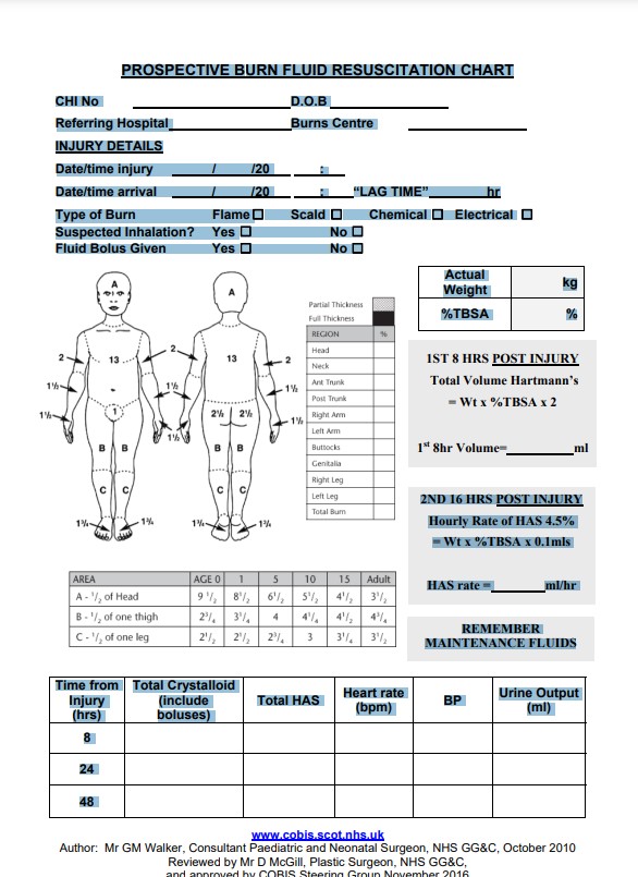
  • The amount of fluid administered should be recorded on the Prospective Fluid Resuscitation Chart along with physiological parameters.

Resuscitation of paediatric burns COBIS guideline

  • Weigh the child on arrival in emergency department
  • Assess and document BSA burn on the Lund & Browder chart provided (simple erythema should not be included)
  • Obtain Intravenous access
  • The initial resuscitation period is 24 hours, split into 2 periods;
    • First 8 hours: modified Parkland formula: 
      • Total volume of Hartmann’s = %TBSA x wt (in kg) x 2 i.e total volume over (8hr – “lag time”)
      • It is a clinical decision whether to include bolus volumes in the total amount.
    • Second 16 hours: 
      • Further fluid given as colloid solution
      • Hourly rate of albumin 4.5% = %TBSA x Wt (in Kg) x 0.1mls
  • In addition to maintenance fluids:
    • 100ml/kg/day for the first 10 kg body weight
    • + 50 ml/kg/day over 10kg and less than 20 kg body weight
    • 20ml/kg/day for each kg over 20kg body weight.
  • Oral fluids are subtracted from maintenance fluids

Suggested observations:

  • Heart rate.
  • Urine output.
  • Blood pressure.
  • Capillary refill.
  • Skin-core temperature gap.
  • Acid base balance.
  • Central venous pressure (if available and indicated)
  • Conscious level.
  • Electrolytes
  • Haematocrit

Prospective burn fluid resuscitation chart

Please chart cumulative totals for fluids and urine output i.e. values from time 0.


References

Alderson P, Bunn F, Li Wan Po A, Li L, Roberts I, Schierhout G. Human albumin solution for resuscitation and volume expansion in critically ill patients. Cochrane Database of Systematic Reviews 2004, Issue 4. Art. No.: CD001208. DOI: 10.1002/14651858.CD001208.pub2.

Arturson G, Jonsson CE. Transcapillary transport after thermal injury. Scand J Plast Reconstr Surg 1979;13:29 –38.

Baker RHJ, Akhavani MK, Jaliali N. Resuscitation of thermal injuries in the United Kingdom and Ireland. Journal of Plastic, Reconstructive and Aesthetic Surgery 2007;60;642-45

Baxter CR, Shires T. Physiological response to crystalloid resuscitation of severe burns. Ann N Y Acad Sci 1968;150: 874 –94.

Blumetti J, Hunt J, Arnoldo B, Parks JK, Purdue GF. The Parkland Formula Under Fire: Is the Criticism Justified? Journal of Burn Care Research 2008;29:180 –186

Cancio LC, Chavez S, Alvarado-Ortega M, et al. Predicting increased fluid requirements during the resuscitation of thermally injured patients. J Trauma 2004;56:404 –13.

Cartotto RC, Innes M, Musgrave MA, Gomez M, Cooper AB. How Well Does The Parkland Formula Estimate Actual Fluid Resuscitation Volumes? J Burn Care Rehabil 2002;23:258 –265

Cochrane Injuries Group Albumin Reviewers. Human albumin administration in critically ill patients: systemic review of randomised controlled trials. Br Med J 1998;317:235–40.

Demling RH. The burn edema process: current concepts. J Burn Care Rehabil 2005;26:207–27.

Demling RH, Kramer GC, Gunther R, Nerlich M. Effect of nonprotein colloid on postburn edema formation in soft tissues and lung. Surgery 1984;95:593– 602.

Engrav LH, Colescott PL, Kemalyan N, et al. A biopsy of the use of the Baxter formula to resuscitate burns or do we do it like Charlie did it? J Burn Care Rehabil 2000;21:91–5.

Gibran NS, Heimbach DM. Current status of burn wound pathophysiology. Clin Plast Surg 2000;27:11–22.

Greenhalgh DG. Burn resuscitation. J Burn Care Res 2007; 28:555– 65.

Guha SC, Kinsky MP, Button B, et al. Burn resuscitation: crystalloid versus colloid versus hypertonic saline hyperoncotic colloid in sheep. Crit Care Med 1996;24:1849 –57.

Holm C, Mayr M, Tegeler J, et al. A clinical randomized study on the effects of invasive monitoring on burn shock resuscitation. Burns 2004;30:798 – 807

Klein MB, Hayden D, Elson C, et al. The association between fluid administration and outcome following major burn: a multicenter study. Ann Surg 2007;245:622– 8.

Lund CC, Browder NC. The estimate of areas of burns. Surgery, Gynecology and Obstetrics 1944;79;352-358. Moore FD. The body-weight burn budget. Basic fluid therapy for the early burn. Surg Clin North Am 1970;50:1249 – 65.

Moore FD. The body-weight burn budget. Basic fluid therapy for the early burn. Surg Clin North Am 1970;50:1249 – 65.

Moyer CA, Margraf HW, Monafo WW Jr. Burn shock and extravascular sodium deficiency—treatment with Ringer’s solution with lactate. Arch Surg 1965;90:799 – 811.

Moylan JA, Mason AD Jr, Rogers PW, Walker HL. Postburn shock: a critical evaluation of resuscitation. J Trauma 1973; 13:354 – 8.

Pham TN, Cancio LC, Gibran NS. American Burn Association Practice Guidelines; Burn Shock Resuscitation. Journal of Burn Care & Research 2008;29(1):257-266

Pruitt BA Jr. Protection from excessive resuscitation: “pushing the pendulum back”. J Trauma 2000;49:567– 8.

Reynolds EM, Ryan DP, Sheridan RL, et al. Left ventricular failure complicating severe pediatric burn injuries. J Pediatr Surg 1995;30:264 –9.

Scott JR, Muangman PR, Tamura RN, et al. Substance P levels and neutral endopeptidase activity in acute burn wounds and hypertrophic scar. Plast Reconstr Surg 2005; 115:1095–102.

Sheridan RL, Tompkins RG, McManus WF, Pruitt BA Jr. Intracompartmental sepsis in burn patients. J Trauma 1994; 36:301–5.

Shires T. Consensus Development Conference. Supportive therapy in burn care. Concluding remarks by the chairman. J Trauma 1979;19:935– 6.

Sullivan SR, Ahmadi AJ, Singh CN, et al. Elevated orbital pressure: another untoward effect of massive resuscitation after burn injury. J Trauma 2006;60:72– 6.

The SAFE Study Investigators. A Comparison of Albumin and Saline for Fluid Resuscitation in the Intensive Care Unit. N Engl J Med 2004;350:2247-56.

Vincent J-L, Navickis RJ, Wilkes MH. Morbidity in hospitalized patients receiving human albumin: A meta-analysis of randomized, controlled trials. Crit Care Med 2004; 32:2029 –2038

Warden GD. Fluid resuscitation and early management. In Total Burn Care. Ed Hendron DN. Publishers Elsevier Health Sciences 2007

Webb J. Current attitudes to burns resuscitation in the UK. Burns 2002;28:205

Wharton SM, Khanna A. Current attitudes to burns resuscitation in the UK. Burns 2001;27:183–4.

Editorial Information

Last reviewed: 30/11/2018

Author(s): Walker, G.M., McGill, D..