1. Right Decisions
  2. Maternity & Gynaecology Guidelines
  3. Maternity
  4. Back
  5. Ultrasound
  6. Transvaginal ultrasound evaluation of the cervix measurement of cervical length (566)
Update your RDS mobile app to v4.7.2 to download toolkits even when website is down.

We are pleased to advise that deep linking capability, enabling users to directly download individual mobile toolkits, has now been released on the RDS mobile app. You will see that each toolkit has a small QR code icon in the header area beside the search icon – see screenshot below. Clicking on this icon will open up a window with a full-size QR code and the alternative of a short URL for sharing with users. Instructions are provided.

You may need to actively update to the latest release - RDS app version 4.7.1 - to see this improvement.

Updating to this latest version of the RDS app is also strongly recommended to get the full benefits of the new resilience  arrangements – specifically, that if the RDS website should fail, you will still be able to download new mobile app toolkits. To check your current RDS version, click on the three dots bottom right of the RDS app screen. This takes you to a “More” page where you will see the version number.  To install latest updates:

On iPhones – go to the Apple store, click on your profile icon top right, scroll down to see the apps waiting to be updated and update the RDS app.

On Android phones – these can vary, but try going to the Google Play store, click on your profile icon top right, click on “Manage apps and device”, select and update the RDS app.

Please get in touch with ann.wales3@nhs.scot with any questions.

Transvaginal ultrasound evaluation of the cervix measurement of cervical length (566)

Warning
Please report any inaccuracies or issues with this guideline using our online form
  1. The probe should be placed in the anterior fornix of the vagina
  2. The maternal bladder should be essentially empty.
  3. A sagittal view of the cervix should be obtained with confidence.
    Remember that the longitudinal axis of the cervix often does not lie exactly within the maternal sagittal axis.
  4. The amount of pressure exerted with the transducer should be kept to a minimum to avoid artificially lengthening the cervix.
  5. The widest viewing angle of the available ultrasound field should be used.
  6. The sonolucent endocervical mucosa should be identified as a guide to the true position of the internal os. The callipers should be placed to measure the linear distance between the triangular area of echodensity at the external os and the V-shaped notch at the internal os.
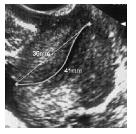
  1. The presence of funnelling should be noted but attempts should not be made at trying to measure it.
  2. The shortest cervical measurement should be recorded rather than an average of the measurements obtained.

Editorial Information

Last reviewed: 10/11/2022

Next review date: 10/11/2027

Author(s): Marie Anne Ledingham.

Version: 2

Approved By: Obstetrics Clinical Governance Group

Reviewer name(s): Donna Bean.

Document Id: 566

References

Sonek j and Shellhaas C. Cervical sonography a review. Ultrasound in Obstetrics and Gynaecology, 1998;11:71-78

To MS, Skentou C, Chan A, Zagaliki A and Nicolaides KH. Cervical assessment at the routine 23 week scan: standardising techniques. Ultrasound in Obstetrics and Gynaecology 201; 17:217-219