1. Right Decisions
  2. Maternity & Gynaecology Guidelines
  3. Maternity
  4. Back
  5. Common obstetric problems, intrapartum labour ward
  6. Umbilical Cord Prolapse (344)
Update your RDS mobile app to v4.7.2 to download toolkits even when website is down.

We are pleased to advise that deep linking capability, enabling users to directly download individual mobile toolkits, has now been released on the RDS mobile app. You will see that each toolkit has a small QR code icon in the header area beside the search icon – see screenshot below. Clicking on this icon will open up a window with a full-size QR code and the alternative of a short URL for sharing with users. Instructions are provided.

You may need to actively update to the latest release - RDS app version 4.7.1 - to see this improvement.

Updating to this latest version of the RDS app is also strongly recommended to get the full benefits of the new resilience  arrangements – specifically, that if the RDS website should fail, you will still be able to download new mobile app toolkits. To check your current RDS version, click on the three dots bottom right of the RDS app screen. This takes you to a “More” page where you will see the version number.  To install latest updates:

On iPhones – go to the Apple store, click on your profile icon top right, scroll down to see the apps waiting to be updated and update the RDS app.

On Android phones – these can vary, but try going to the Google Play store, click on your profile icon top right, click on “Manage apps and device”, select and update the RDS app.

Please get in touch with ann.wales3@nhs.scot with any questions.

Umbilical Cord Prolapse (344)

Warning
Please report any inaccuracies or issues with this guideline using our online form

Umbilical cord prolapse is an obstetric emergency that complicates 0.1 – 0.6% of all deliveries (1% if breech). One major study found perinatal mortality rate of 91/1000.1 Within a hospital setting the majority of fetal morbidity is linked with prematurity and congenital malformations however birth asphyxia is also a cause of morbidity associated with cord prolapse.

Definitions

Umbilical Cord Prolapse = a loop of cord below the presenting part with ruptured membranes whether visible or felt on examination

Umbilical Cord Presentation = a loop of cord below the presenting part with intact membranes.

In practical terms acute management is identical and should be immediate.

Risk factors

The majority of cases of umbilical cord prolapse are seen in women at term with babies of normal birth weight and cephalic presentation however table 1 highlights several clinical features which increase the risk of this obstetric emergency:

Table 1. Risk factors for cord prolapse

General

Procedure related

Multiparity

Artificial rupture of membranes

Low birth weight, less than 2.5kg

Vaginal manipulation of the fetus with ruptured membranes

Prematurity less than 37 weeks

External cephalic version (during procedure)

Fetal congenital anomalies

Internal podalic version

Breech presentation

Stabilising induction of labour

Transverse,  oblique  and  unstable  lie  (when the longitudinal axis of the fetus is changing repeatedly)

Insertion of uterine pressure transducer

Second twin

Polyhydramnios

Unengaged presenting part

Low-lying placenta, other abnormal placentation

Prevention

  • Awareness of Risk Factors. Inform patient of risk if recognised risk factor.
  • With transverse, oblique or unstable lie elective admission after 37+0 should be discussed and women in the community should be advised to present urgently if signs of labour or suspicion of membrane rupture.
  • Always perform an abdominal examination and determine the fetal lie is longitudinal. Only perform forewater amniotomy if you are confident that the head is in the pelvis. If ARM is necessary with a high presentating part ensure there is access to facilities for immediate CS
  • Avoid upward pressure on the presenting part during VE, FBS, ARM.
  • Women with non-cephalic presentation and PPROM should be recommended to have inpatient care.
  • Routine ultrasound examination is not sufficiently sensitive or specific for identification of cord presentation antenatally and should not be performed to predict increased probability of cord prolapse, unless in the context of a research setting.
  • Sensitive ultrasound screening can be considered for women with breech presentation at term who are considering vaginal birth attempt.

Management

Recognition:

  • Cord presentation and prolapse may occur without outward physical signs and with a normal fetal heart rate pattern. Exclude palpable cord at every vaginal examination in labour and after spontaneous rupture of membranes if risk factors are present or if CTG abnormalities commence soon thereafter.
  • Cord prolapse should be suspected where there is an abnormal fetal heart rate pattern (bradycardia, variable decels etc), particularly if such changes commence soon after membrane rupture, spontaneously or with amniotomy.
  • Speculum and/or digital vaginal examination should be performed when cord prolapse is suspected.
  • If no cord pulsation, ultrasound scan should be used to confirm fetal viability. Fetal heart movements may be visualised by ultrasound scan in the absence of cord pulsation.
  • With spontaneous rupture of membranes in the presence of a fixed cephalic presentation, normal fetal heart rate patterns and the absence of risk factors for cord prolapse, routine vaginal examination is not indicated if the liquor is clear.

Call for Help:

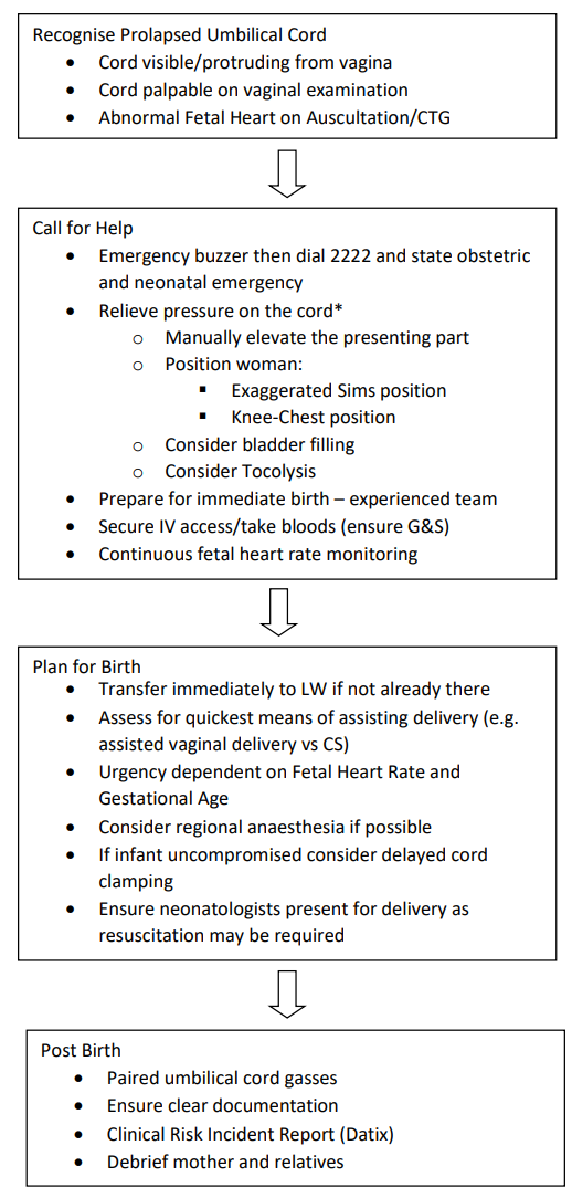
  • Activate emergency buzzer to call for assistance. 2222 call and state obstetric and neonatal emergency.
  • If cord prolapse happens out of hospital an emergency ambulance should be called immediately to transfer woman to the nearest consultant-led obstetric unit. Even if birth appears imminent, an ambulance should be called in case of neonatal compromise. Liaise with obstetric unit and inform of emergency and estimated time of arrival.

Relieve Pressure on the Cord;

  • As soon as cord prolapse is recognised cord compression should be minimised by elevating the presenting part.
  • Maternal positioning:
    • Knee-chest, face-down position is traditionally recommended however this is not suitable for ambulance transfer.
    • Exaggerated Sims position (left-lateral with a pillow under the left hip) with or without Trendelenberg (tilted bed so that the woman’s head is lower than her pelvis) may be used instead.
  • Digital Elevation of the Presenting Part
    • Clinician’s gloved fingers should be kept within the vagina to elevate the presenting part.
    • If the cord has prolapsed out of the vagina attempt to gently replace it back into the vagina using a dry pad and with minimal handling. Any handling of the cord can cause vasospasm.
    • Do not attempt to replace cord above the presenting part.
    • There is no evidence to support the practice of covering the exposed cord with sterile gauze soaked in warmed saline.
  • Bladder Filling
    • If decision to birth interval is likely to be long e.g. due to hospital transfer elevating the presenting part through bladder filling may be considered.
    • Insert Foley catheter into the urinary bladder and allow urine to drain. Fill bladder with sterile 0.9% NaCL using an intravenous infusion set. The catheter should be clamped once 500ml has been instilled.
    • Ensure bladder is emptied prior to any method of birth being attempted.
  • Reduce Contractions
    • Stop syntocinon infusion immediately if running
    • Consider tocolysis (Terbutaline 0.25mg s/c) to reduce contractions and improve fetal bradycardia when there is a cord prolapse.

Plan for Birth

  • Immediate transfer to LW
  • Ensure IV access in place and up to date FBC, G&S taken.
  • Assessment for birth:
    • If the cervix is not fully dilated, caesarean section should be performed.
      • A category 1 CS should be performed with aim to deliver within 30 minutes but without compromising maternal safety if there is evidence of fetal heart rate abnormalities. Verbal consent is sufficient.
      • Category 2 caesarean section can be conducted in women in whom the fetal heart rate is normal, but continuous assessment of fetal heart trace is essential. If CTG becomes abnormal recategorisation to category 1 should be immediately considered.
    • If the cervix is full dilated, consider an operative vaginal birth as long as it is anticipated that it would be accomplished quickly and safely.
      • Ventouse or forceps should only be considered if the prerequisites for operative vaginal birth are met.
  • Delayed cord clamping may be considered as long as the baby is uncompromised.
  • Breech extraction may be performed under some circumstances, for example after internal podalic version for the second twin.
  • Prolonged or repeated attempts at regional anaesthesia should be avoided.
  • An experienced neonatal team must be present at birth to ensure full cardiorespiratory support is given to the neonate, if required.

Post Birth

  • Ensure paired umbilical cord gasses are taken
  • Clear documentation of events and times of each method used to alleviate pressure on the cord.
  • Complete an incident reporting form (Datix)
  • Debrief patients on events.

Management at the threshold of viability (23+0 – 24+6)

  • If time: discuss the woman and neonatologists about outcomes for baby and risks to the mother from any interventions.
  • Expectant management should be discussed for cord prolapse complicating pregnancies with gestational age at the threshold of viability.
  • Women should be counselled on both continuation and termination of pregnancy following cord prolapse at the threshold of viability.

Adapted from RCOG Green Top Guideline 50 and PROMPT Cord Prolapse.

Editorial Information

Last reviewed: 01/06/2022

Next review date: 30/06/2024

Author(s): Dr Julie Murphy.

Version: 3

Approved By: Obstetrics Clinical Governance Group

Document Id: 344

References
  1. Murphy DJ, MacKenzie IZ. The mortality and morbidity associated with umbilical cord prolapse. Br J Obstet Gynaecol 1995;102:826–30.
  2. Royal College of Obstetricians and Gynaecologists. Umbilical Cord Prolapse. Green-top Guideline No. 50. London: RCOG;2014 
  3. Module 11. Cord Prolapse. Practical Obstetric Multi-Professional Training. PROMPT. 3rd Cambridge: 2019. p217-227.