1. Right Decisions
  2. GGC - Clinical Guidelines
  3. Maternity
  4. Back
  5. Antenatal, general
  6. Invasive Placental Disease (577)
Please update your RDS mobile app to version 4.7.1

We are pleased to advise that deep linking capability, enabling users to directly download individual mobile toolkits, has now been released on the RDS mobile app. When you install the update, you will see that each toolkit has a small QR code icon the header area beside the search icon – see screenshot below. Clicking on this icon will open up a window with a full-size QR code and the alternative of a short URL for sharing with users. Instructions are provided.

You may need to actively install the update to install RDS app version 4.7.1 to see this improvement. Installing this update is also strongly recommended to get the full benefits of the new contingency arrangements – specifically, that if the RDS website should fail, you will still be able to download new mobile app toolkits. 

To check your current RDS version, click on the three dots bottom right of the RDS app screen. This takes you to a “More” page where you will see the version number.  To install latest updates:

On iPhones – go to the Apple store, click on your profile icon top right, scroll down to see the apps waiting to be updated and update the RDS app.

On Android phones – these can vary, but try going to the Google Play store, click on your profile icon top right, click on “Manage apps and device”, select and update the RDS app.

Please get in touch with ann.wales3@nhs.scot with any questions.

Invasive placental disease (577)

Warning

Objectives

Please report any inaccuracies or issues with this guideline using our online form

This guideline covers the diagnosis and subsequent management (antenatal and intrapartum of women with suspected abnormally invasive placental disease in GG&C

There should be a suspicion of this condition in all women with a previous section and a low-lying placenta in a subsequent pregnancy. It requires a multi-disciplinary management team and is frequently associated with heavy bleeding, hysterectomy and complex antepartum, intrapartum and postpartum morbidity. Senior staff should be involved in all decisions. Careful antenatal planning and good communication are vital in the management of these women.

This guideline should be used in conjunction with the AIP checklist to minimise the chance of avoidable errors.

Diagnosis

Ultrasound

Undiagnosed cases are associated with more complications and significant morbidity; a high level of suspicion in women with previous sections and a low anterior placenta is required. Women with other uterine surgery should also prompt careful assessment of the placenta. These operations may be elsewhere in the uterus.

Risk Factors:

  • Caesarean section
  • Myomectomy
  • Hysterotomy
  • Endometrial Ablation
  • Uterine Perforation

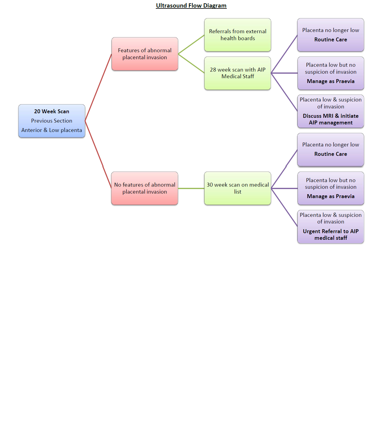
20 week ultrasound may show anterior low placenta with a previous section. If there are no suspicious features then repeat sonographer scan at 30 weeks. If placenta remains anterior and low at 30 weeks with or without features suspicipous of invasion a medical scan should be arranged.

If suspicious features are already present then a 28 week scan with a medical AIP sonographer should be arranged. See the flow chart below for management plans.

It is anticipated that referrals from external health boards may present at this stage. These women should have been scanned by a medical sonographer in their own unit and be referred directly to one of the AIP consultants at around 30 weeks. One of the AIP medical staff will then arrange an ultrasound scan to assess their placenta.

In women with a confirmed ultrasound diagnosis further imaging with MRI can provide useful information to help plan surgery. Please see the flowchart and MRI criteria in this document.

Abnormally Invasive Placenta - Ultrasound Assessment Form

Magnetic Resonance

  1. A referral for MRI of the placenta (TRAC request MR Obstetric) should be made only after a possible or probable risk of AIP is raised by one of the AIP medical sonographers triggering entry of that patient onto the AIP pathway. This referral for MRI will probably be at 30 weeks.
  2. The referral for MRI should be to a named radiologist at the site of referral; they should also be contacted by e-mail- see list below
  3. The referral information should include
    • A link to USS images if possible and/or a list of the specific, positive USS findings (copy of USS proforma shown in guideline included in request or on Portal). This information should include the site of concern and totalled estimated risk score and features on ultrasound.
    • EDD, parity, number of previous LUSCS.
    • Consideration of MRI contraindications.
  1. An MRI scan will be appointed at the site of referral.
  2. The MRI scan can be performed to the agreed local GGC imaging protocol and will ideally be reviewed and reported by 2 radiologists.
  3. MRI is adjunctive and complementary to USS and should not be interpreted or acted upon in isolation.
  4. MDT discussion for patients on the AIP pathway is advised to discuss the salient USS and MRI imaging findings with the surgical, obstetric and pathology team to guide subsequent management and interpretation.
  5. Surgical, IR, pathological and medical photography feedback to radiology in ALL cases in the first instance to Emily.Stenhouse@ggc.scot.nhs.uk

If the diagnosis of AIP is confirmed or seems likely then progress to the management section; otherwise manage as other placenta praevia cases. (see praevia guideline )

Antenatal management

Commence iron therapy following diagnosis. Inform one of the named invasive placental disease obstetricians (Marcus McMillan, Dawn Kernaghan). They will then organise delivery in main theatres with appropriate equipment and staff (see management flowchart and bundle).

Delivery will usually be between 35 and 36 weeks and performed in the main theatre suite in GRI. In patient admission is recommended from 34 weeks. If admitted:

  • Valid group & save at all times
  • IV access is NOT required unless bleeding or unwell
  • Complete invasive placental disease bundle as soon as feasible
  • Inform clinical psychology of admission
  • Appropriate weight-based LMWH (Low Molecular Weight Heparin) prophylactic dose
    • The final dose of LMWH should be the day prior to surgery; i.e. if surgery planned for a Wednesday the final dose is on Tuesday. Discussing timing with consultant anaesthetist

If admission is declined then careful counselling is required emphasising:

  • Proximity to hospital
  • Constant companion
  • Clear plan for what to do if bleeding starts
  • Admission day prior to surgery as a MINIMUM
  • Risk of catastrophic haemorrhage

There should be a clear plan for delivery documented. This should include the planned incision (midline or transverse) along with the expectation of placental delivery, uterine resection or hysterectomy. Additional specialties required should be documented. It is important to remember that the expected plan may change upon opening the abdomen and that any antenatal discussions regarding delivery should reflect this.

A theatre date should be set by the lead obstetrician for the case following discussion with the other members of the team. This will include (but is not limited to):

  • Gynaecology oncology consultant
  • Anaesthetic consultant
  • Interventional radiology consultant
  • Urological consultant
  • Management representative (Vicki Mazzoni)
  • Labour ward theatre coordinator
  • Main Theatres manager

Consideration needs to be given to other planned obstetric workload and appropriate staffing arrangements made. Named contacts for each of these specialties are provided in the contact details section of this guideline; these people may not necessarily be at the surgery but would arrange appropriate cover from their specialty.

Once a date is set the woman’s details should be sent to Lea Ferguson (paediatric secretary) so she can be added to the high risk meeting for discussion with the paediatricians. The haematology department should be informed of the planned surgery. Medical photography should be asked to attend.

The referring consultant should be included in communication and informed of the planned delivery date as they may wish to attend the operation; this should be facilitated.

Consent

For the delivery and subsequent obstetric management this should be performed by the responsible consultant obstetrician once patient admitted to hospital.

Consultants from other specialties who will be performing planned procedures should complete their own consent prior to the day of surgery.

The consent form in the checklist includes the following risks:

  • Haemorrhage, transfusion and cell salvage
  • Infection
  • Thromboembolism
  • Midline laparotomy
  • Hysterectomy with conservation of ovaries and loss of fertility
  • Resection of adherent myometrium
  • Bladder and ureteric injury and repair
  • Bowel injury
  • Maternal death
  • Neonatal death
  • ICU admission
  • Return to theatre
  • Post-operative pain and prolonged recovery
  • Fetal lacerations
  • Neonatal unit admission

Consent for medical photography should be obtained as this is a useful adjunct for review of these cases in conjunction with MRI and ultrasound scans. A separate consent form is required for this. Similarly consent for pathology specimen use should be obtained.

Repeat ultrasound assessment of the placenta by one of the invasive placental disease consultants (Marcus McMillan, Dawn Kernaghan) should be undertaken during the in-patient stay. This can be useful for planning management including abdominal incision and likelihood of placental delivery.

Surgical Planning

It is anticipated that the vast majority of women will require common iliac prophylactic balloons from interventional radiology. If this is not to be undertaken it should be agreed with the entire surgical team and reasoning clearly documented. Placing balloons in an emergency may not be appropriate or possible.

Ureteric stenting should be considered routine in all women with a planned or likely hysterectomy. In women with a planned excisional or conservative approach they may still be of use and there should be discussion with the surgical team on the appropriate course. Again, these will not be feasible to site in an emergency situation arising before or during an operation.

There should be an idea of the planned surgery before it is undertaken. Points to be considered include:

  • Should there be an attempt to deliver the placenta?
  • Is resection an option for this case?
  • Will conservative management be required?
  • What are the woman’s plans for future fertility?

The intent with resection or conservative management is NOT to preserve fertility but to minimise the surgical risk to the patient. In these situations sterilisation should be discussed. If sterilisation is declined ensure the risks of recurrent AIP in a future pregnancy are discussed and documented.

Clinical Psychology

There are significant psychological issues to consider with these cases, many women unsurprisingly find the experience very traumatic:

  • Prolonged and imposed hospital admission away from family
  • Significant risk of severe morbidity and death
  • Complex surgery with large scar
  • Enforced sterility
  • Protracted recovery

As such input from the clinical psychology department can play a valuable role in preparing these women for coping with their delivery and in their recovery afterwards. While some women may decline input at the very least clinical psychology should be made aware of them.

Visiting Consultants

Referring obstetric consultants or trainees from other hospitals may be keen to attend for these cases. This should be facilitated whenever possible however the total number of people in theatre should be limited to 3 trainees across all specialties.

Day of surgery

A request for 4 units of crossmatched blood should be made

Aim for patient leaving ward 08:30

Team brief in theatre with all members present:

  • Obstetrics
  • Gynaecology
  • Anaesthetics
  • Urology
  • Interventional Radiology
  • Scrub Nurse
  • Midwifery (to receive baby)
  • Operating Department Practitioners & Anaesthetic Assistants
  • Theatre Nursing Staff
  • Interventional Radiographers
  • Health Care Assistants

The planned procedure should be discussed along with the expected timeline and anticipated difficulties. Everyone should be clear on their role and responsibilities. Theatres can easily become very noisy and busy with a big team; clear communication in these cases is vital so quiet is requested during the brief. Those without a need to be present will be asked to leave.

The anticipated order of procedures is usually:

  • Spinal anaesthetic (if appropriate)
  • Cystoscopy and ureteric stenting
  • Common iliac balloon placement
  • General anaesthetic & Laparotomy

Before general anaesthetic:

  • Surgical pause
  • Ensure neonatal staff available
  • Confirm all appropriate staff present
  • Inform medical photography of operation starting

Neonatal Attendance

Staff & equipment required (minimum):

Neonatal nurse + middle grade trainee confident in airway management Transport incubator + kit bag

Schedule:

Neonatal nurse to attend whilst theatre being prepared:

  • Resuscitaire set up and checked
  • ET tube available (3.5mm tube if term delivery) + Guedel airway size 0
  • Transport incubator on and plugged in         
  • Kit bag including naloxone

Neonatal nurse can then return to SCBU

Middle grade trainee made aware of impending call. Ensure neonatal nurse familiar with correct pager number (12200)

Theatre team to call 25247 as patient draped

Neonatal nurse pages trainee and both attend immediately

Operation

During the operation it is easy to be come very focussed on a particular task. Good communication in these cases is essential and it is important that information is freely shared around the theatre.

Intraoperative documentation should be performed by a designated scribe with particular regard to use of blood products, EBL and cell salvage.

Medical photography is a useful tool for case review and patient debrief however clinical judgement should be used when pausing for photographic opportunities.

A “sign out” should be completed at the end of the procedure including as a minimum: Swab count, instrument check, sharps check, EBL, post-operative plan, analgesia plan, thromboprophylaxis plan, specimens for pathology.

The operation notes should be completed by all the involved surgeons. An appropriate pathology request should be completed. A copy of the operation findings and the pathology report should be sent to Emily Stenhouse.

Reflection, Debrief and Data

The team should debrief together after the case and reflect on what went well and any potential areas for improvement. Relevant learning points can be incorporated into future cases; it is the role of the consultant obstetrician to advance this.

These are complex cases with high risks of complications. Mistakes and problems will be encountered and it is important to identify and address these along with potential learning issues.

The service is one that will be consistently improving. Gathering good quality data will facilitate this. The obstetric and anaesthetic consultants involved should ensure the checklist is appropriately completed and ensure these cases are entered into the AIP database.

Postnatal management

Initially this will likely be in labour ward HDU or possibly ICU depending on the clinical situation.

Daily consultant review is required until fit for discharge. Women in the Rooming-In Suite do not receive regular obstetric review and should not be admitted there until they are fit to leave the maternity hospital.

Many of the women having a caesarean hysterectomy have remarked on how difficult they have found the transition to the postnatal wards. It should be remembered that they have not had a “normal” caesarean section and may have more dependency than other patients.

Going Home

6 weeks of LMWH is required.

A follow-up appointment at PRM to discuss events once pathology is available should be arranged. The obstetric consultant will arrange this appointment.

Ensure that clinical psychology have been made aware of the patient.

These women are at high risk of postnatal complications. It should be clearly explained to them to seek medical advice if they have concerns. There should be a clear channel of communication for them to contact PRM if they have questions or concerns.

Emergency presentation

Many of these cases will present as emergencies. When mothers are unstable or compromised management may differ from uncompromised women delivered for fetal indications. Phoning consultant colleagues for assistance in these situations is entirely appropriate and is encouraged.

In unstable women prompt recourse to emergency section and hysterectomy may be life saving. CONSULTANT obstetricians, gynaecologists and anaesthetists should be involved as quickly as possible.

Any woman with suspected invasive placental disease where delivery is being considered should be seen by the on-call obstetric consultant. The decision for delivery should be made and undertaken by a consultant unless the woman presents in extremis; the decision to deliver should not be left until this point. If there is catastrophic haemorrhage then fetal wellbeing is not an immediate concern and management of the mother should ALWAYS take priority.  Due to the complex nature of these cases the on-call gynaecology consultant and anaesthetic consultant should also attend.

Crossmatch 4 units of blood immediately. A midline laparotomy extending from symphysis beyond the umbilicus will be required. The goal is to make a high transverse uterotomy to avoid the placenta. If time allows then ultrasound assessment of the placental upper margin could be performed and an appropriate midline incision made. Once the baby has been delivered then assessment of the placenta can be made.

  • If there is maternal compromise then hysterectomy may be required. This should not be delayed if it is felt the placenta cannot or should not be delivered or if bleeding cannot be arrested.
  • If delivery is for fetal indications and there is no bleeding then the uterus and abdomen can be closed and definitive management undertaken once consultant colleagues and measures such as cell salvage and interventional radiology are available.
  • Urological input may be required; the on-call consultant should be contacted through switchboard if needed.
  • Interventional radiology may be of benefit however in cases where blood loss has causedmaternal compromise it may not be appropriate to delay surgery for this reason.
  • Attempted delivery of the placenta may cause or exacerbate bleeding; careful consideration should be given before undertaking this.

See also the Major Obstetric Haemorrhage guideline

Useful Contacts

Name

Role

E-mail

Marcus McMillan

Obstetrics

Marcus.McMillan@ggc.scot.nhs.uk

Dawn Kernaghan

Obstetrics

dawn.kernaghan@ggc.scot.nhs.uk

Ann Duncan

Obstetrics

Ann.Duncan@ggc.scot.nhs.uk

Kevin Burton

Gynaecology Oncology

Kevin.Burton@ggc.scot.nhs.uk

Rhona Lindsay

Gynaecology Oncology

Rhona.Lindsay@ggc.scot.nhs.uk

Andrew Christie

Interventional Radiology

Andrew.christie@ggc.scot.nhs.uk

Ram Kasthuri

Interventional Radiology

Ram.Kasthuri@ggc.scot.nhs.uk

Michael Palmer

Urology

Michael.palmer@ggc.scot.nhs.uk

Mary Brown

Urology

Mary.brown3@ggc.scot.nhs.uk

Drew Smith

Anaesthetics

Drew.smith@ggc.scot.nhs.uk

Malcolm Broom

Anaesthetics

Malcolm.Broom@ggc.scot.nhs.uk

Niels Weidenhammer

Anaesthetics

Niels.weidenhammer@ggc.scot.nhs.uk

Emily Stenhouse

Radiology

Emily.stenhouse@ggc.scot.nhs.uk

Chris Lilley

Neonates

Chris.Lilley@ggc.scot.nhs.uk

Catherine Bagot

Haematology

Catherine.bagot@ggc.scot.nhs.uk

Carol Fergusson

Senior Charge Midwife

Carol.fergusson@ggc.scot.nhs.uk

Rebecca Hutchison

Senior Charge Midwife

Rebecca.hutchison@ggc.scot.nhs.uk

John Ruth

Cell Salvage

John.ruth@ggc.scot.nhs.uk

Annemarie McNally

Anaesthetic Nurse

Annemarie.mcnally@ggc.scot.nhs.uk

Kirsten McKillop

Psychology

Kirsten.mckillop@ggc.scot.nhs.uk

Alison Robertson

Psychology

Alison.robertson2@ggc.scot.nhs.uk

Janet Cheaitou

Theatre Manager

Janet.cheaitou@ggc.scot.nhs.uk

Stephanie Leca

Theatre coordinator

Stephanie.leca@ggc.scot.nhs.uk

Katie McEwan

Clinical Services Manager

Katie.mcewan@ggc.scot.nhs.uk

Vicki Mazzoni

Lead Midwife

Vicki.mazzoni@ggc.scot.nhs.uk

Claire Stewart

Clinical Services Manager

Claire.stewart@ggc.scot.nhs.uk

Lea Ferguson

Neonates Secretary

Lea.ferguson@ggc.scot.nhs.uk

Dawn Penman

Pathology

Dawn.penman@ggc.scot.nhs.uk

Urology Secretaries         

25493

25492

Urology pager                    

13278

Radiology Secretaries 

24619

Obstetric Secretaries 

25291

Medical Photography 

24692

20510

Editorial Information

Last reviewed: 10/08/2017

Next review date: 01/09/2022

Author(s): Marcus McMillan.

Approved By: Obstetrics Clinical Governance Group

Document Id: 577