Key questions

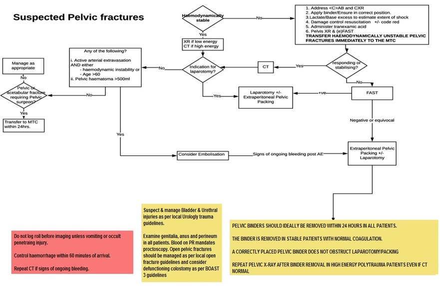
  • Is the patient haemodynamically stable?
  • Is the pelvic fracture mechanically stable?
  • Are there other sites of bleeding including chest and abdomen?
  • Is the fracture open? (perineum, rectum, vagina)?

Pelvis diagram

Principles

Mortality of up to 35% is reported with pelvic bleeding. Haemorrhage control should be achieved within 60 minutes of the patient’s arrival.

Most haemorrhage (>70%) is venous and can be controlled by:

  • Minimising patient movement
  • A pelvic binder applied over the greater trochanters
  • Binding the knees and ankles together
  • The principles of damage control resuscitation
  • Arterial bleeding is controlled by four methods:
    • Embolisation
    • Extra-peritoneal Pelvic Packing
    • Thoracotomy with Aortic Cross Clamping.

Log-rolling

  • DO NOT log roll before imaging unless:
    • An occult penetrating injury is suspected in an unstable patient
    • It is required to clear the airway.
  • Log rolling can disrupt clot and cause haemodynamic instability.

Transfer

Transfer to the MTC patients with:

  • haemodynamic instability from a pelvic or acetabular fracture - immediately
  • A failed closed reduction of a native hip joint, where local expertise is unavailable - immediately
  • Pelvic and acetabular fractures who require specialist pelvic reconstruction - <24 hrs from injury.

Imaging

X-Ray

X-ray is Indicated in the following:

  • Code Red patients
  • Low Energy (Suspected pelvic fractures).

eFAST

eFAST is used in pelvic fracture patients deemed too unstable for CT.

  • A positive FAST scan mandates a laparotomy after pelvic external fixation (if indicated by fracture pattern) followed by Extra-Peritoneal Pelvic Packing (EPP).
  • A negative or equivocal FAST suggests Extra-Peritoneal Pelvic Packing (EPP) after pelvic external fixation (if indicated by fracture pattern) followed by laparotomy.

CT

Use CT for suspected high-energy pelvic fractures.

X-ray machine
Image credit: Ben Stephenson from Beltsville, Maryland, CC BY 2.0

Embolisation

Keep the anterior pelvic peritoneum intact.

The absence of extravasation does not exclude pelvic haemorrhage. Consider repeating triple phase CT if clinical signs of ongoing bleeding.

Indications:

  • Active extravasation and hemodynamic instability with no indication for laparotomy.
  • Active extravasation in patients aged>60 with open book, butterfly, or pelvic shear fractures, regardless of haemodynamic stability.
  • Active extravasation regardless of haemodynamically stability, especially in the elderly.

Relative Indications:

  • Active extravasation regardless of haemodynamic stability, especially in the elderly
  • Pelvic haematoma >500ml.

Surgery and extra-peritoneal pelvic packing (EPP)

The pelvic binder (or pelvic external fixator) should remain on during laparotomy and EPP. If a laparotomy is required, perform this first.

A pelvic retroperitoneal zone III injury from blunt trauma should not be explored from within the abdomen.

Extra-Peritoneal Packing from a supra-pubic incision is indicated in the following patients:

  • Patients who require a laparotomy for abdominal injuries
  • Patients in extremis (sustained SBP<70mmHg)
  • Patients in whom IR is not available within 30 minutes
  • Persisting bleeding after embolisation.

Pelvic Binders

Pelvic Binders are a resuscitation aid and should be removed once resuscitation is complete in patients who are normothermic with no further bleeding and normal coagulation. This is usually within 24 hours of admission.

Mechanically unstable pelvic fractures requiring fixation

External Fixation

Consider external fixation in patients undergoing laparotomy and/or EPP with deranged physiology and those receiving damage control surgery only after discussion with a specialist pelvic surgeon.

Internal fixation

Should be performed within 24 hours in stable patients without deranged physiology Patients with deranged physiology should have definitive fixation delayed.

Open pelvic fractures

The lower abdomen, groin, buttocks, perineum, anus, rectum, and vagina must be examined in all cases for wounds. This can be performed intra-operatively or after imaging as appropriate.

A positive PR exam for blood mandates a proctoscopy. A positive PV exam for blood mandates a colposcopy.

Debridement should occur:

  • Immediately if contaminated with agriculture, aquatic or sewage material
  • Within 12 hours if high energy
  • Within 24 hours if low energy.

Wounds to the anus and rectum, and some buttock and perineum wounds often require a defunctioning stoma and general surgery input. The principles of which are:

  • Consider each case carefully on own merits regarding need and timing.
  • Do not perform during damage control.
  • Obtain consent where possible.
  • May be created laparoscopically (for a defunctioning colostomy after distal injury).
  • It should be sited in the upper abdomen remote form the site of pelvic fixation.
  • Rectum/colon injuries from pelvic fractures should be treated with resection or repair, defunctioning stoma and irrigation of the distal bowel segment and pelvic drainage.

Algorithm for suspected pelvic fractures

Suspected pelvic fractures algorithm