Right Decision Service newsletter: October 2024
Welcome to the Right Decision Service (RDS) newsletter for October 2024.
1.Contingency arrangements for RDS outages
Development of the contingency solutions to maximise RDS resilience and minimise risk of future outages is in progress, aiming for completion by Christmas. As a reminder, these contingency arrangements are:
- Optimising mobile app build process
- Mobile app always to be downloadable.
- Serialising builds to mobile app; separate mobile app build from other editorial and end-user processes
- Load balancing – provides failover (also enables separation of editorial processes from other processes to improve performance.)
In the meantime, a gentle reminder to encourage users to download essential clinical toolkits to their mobile devices so that there is an offline version always available.
2. New deployment with improvements.
A new scheduled deployment with minor improvements drawn from support tickets, externally funded projects, information related to outages, and feature requests will take place in early December. Key improvements planned are:
- Deep-linking to individual toolkits within the RDS mobile app. Each toolkit will now have its own direct URL and QR code, both accessible from the app. These can be used to download the toolkit directly where users already have the RDS app installed. If the user does not yet have the RDS app installed, they will be taken to the app store to install the app and immediately afterwards the toolkit will automatically open and download. Note that this will go live a few days later than the improvements below due to the need to link up the mobile front end to the changes in the content management system.
- Introducing an Announcement Header field to replace the hardcoded "Announcements and latest updates" text. This will enable users to see at a glance the focus of new announcements.
- Automated daily emptying of the recycling bin (with a 30 day rolling grace period) in the content management system. A bug preventing complete emptying of the recycling bin contributed to one of the outages earlier this year.
- Supporting multiple passcodes (ticket 6079)
- Expanding accordion section to show location of a search result rather than requiring user coming from a search result to manually open all sections and search again for the term.
- Displaying first accordion section Content text as a snippet on the search results page as a fallback if default/main content is not provided
- Displaying the context of each search result in the form of a link to the relevant parent tool/section. This will help users to choose which search result is most likely to be appropriate for their needs.
- As part of release of the new national benzodiazepine quality prescribing guidance toolkit sponsored by Scottish Government Effective Prescribing and Therapeutics, a digital tool to support creation of benzodiazepine tapering/withdrawal schedules.
We are also seeking approval to use the NHS Scotland logo and title for the RDS app on the app stores to help with audience engagement and clarity around the provenance of RDS.
3. RDS Search, Browse and Archive/Version control enhancements
We are still hopeful that user acceptance testing for at least the Search and browse enhancements can take place before Christmas. Thank you for your patience and understanding in waiting for these improvements. Timescales have been pushed back by old app migration challenges, work to address outages, and most recently implementing the contingency arrangements.
4. Support tickets
We are aware that there continue to be some issues around a number of RDS support tickets, in part due to constraints around visibility for the RDS team of the tickets in the existing support portal. We are investigating the potential to move to a new support ticket requesting system from early in the new year. We will organise the proposed webinar around support ticket processes once we have confirmed the way forward with the system.
Table formatting
There is a known issue with alterations in formatting of some RDS tables which seems to have arisen as a result of the 17 October deployment. Tactuum is working on a fix and on implementing additional regression testing to prevent this issue recurring.
5. New RDS toolkits
Recently launched toolkits include:
NHS Lothian Infectious Diseases
Scottish Health Technologies Group – Technology Assessment recommendations
NHS Tayside Anaesthetics and Critical Care projects – an innovative toolkit which uses PowerAutomate to manage review and response to proposals for improvement projects.
If you would like to promote one of your new toolkits through this newsletter, please contact ann.wales3@nhs.scot
A number of toolkits are expected to go live before Christmas, including:
- Focus on dementia
- Highland Council Getting it Right for Every Child
- Dumfries and Galloway Adult Support and Protection procedures
- National Waiting Well toolkit
- Fertility Scotland National Network
- NHS Lothian postural care for care homes
6.Sign up to RDS Editors Teams channel
We have had a good response to the recent invitation to sign up to the new Teams channel for RDS editors. This provides a forum for editors to share learning, ideas and questions and we hope to hold regular webinars on topics of interest. The RDS team is in the process of joining participants to the channel and we’d encourage all editors to take part, using the registration form – available in Providers section of the RDS Learning and Support area.
7. Evaluation projects
The RDS team has worked with colleagues in NHS Grampian and the Digital Health & Care Innovation Centre to evaluate the impact of the Prevent the progress of diabetes web and mobile app in a small-scale pilot project. This app provides access to local and national resources and services targeted at people with prediabetes, a history of gestational diabetes, or candidates for remission. After just 8 weeks of using the app, 94% of patients reported increased their knowledge and understanding of diabetes, and 88% said it had increased their confidence and motivation to make lifestyle changes, highlighting specific behaviour changes. The learning from this project is informing development of a service model based on tailored support for patient groups with, high, medium and low digital self-efficacy.
Please contact ann.wales3@nhs.scot if you would like to know more about this project.
-
Training sessions for new editors (also serve as refresher sessions for existing editors) will take place on the following dates:
- Friday 29th November 3-4 pm
- Thursday 5 December 3.30 -4.30 pm
To book a place, please contact Olivia.graham@nhs.scot, providing your name, organisation, job role, and level of experience with RDS editing (none, a little, moderate, extensive.)
To invite colleagues to sign up to receive this newsletter, please signpost them to the registration form - also available in End-user and Provider sections of the RDS Learning and Support area. If you have any questions about the content of this newsletter, please contact his.decisionsupport@nhs.scot If you would prefer not to receive future newsletters, please email Olivia.graham@nhs.scot and ask to be removed from the circulation list.
With kind regards
Right Decision Service team
Healthcare Improvement Scotland
The Right Decision Service: the national decision support platform for Scotland’s health and care
Website: https://rightdecisions.scot.nhs.uk Mobile app download: Apple Android

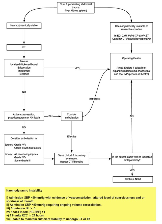
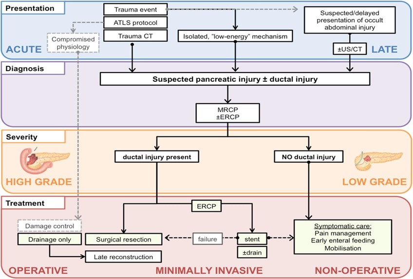
 North of Scotland major trauma guidelines