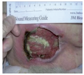
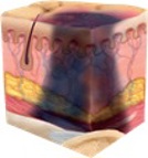
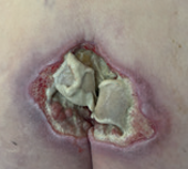
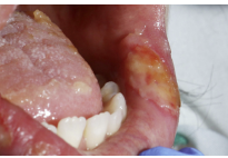
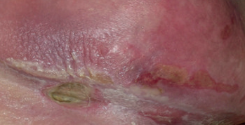
Scottish adaptation of the European Pressure Ulcer Advisory Panel (EPUAP) pressure ulcer classification tool

Warning

Bruising can indicate deep tissue injury.

The depth of a Grade 3 or 4 pressure ulcer varies by anatomical location. Areas such as the bridge of the nose, ears, occiput and malleolus do not have fatty tissue so the depth of these ulcers may be shallow.

This pressure ulcer classification tool is also available in PDF format.

Early warning signs - Blanching/erythema

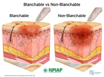
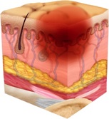
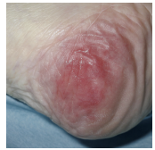
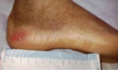
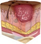
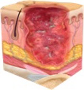
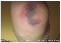
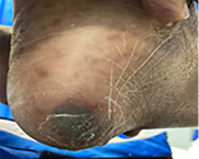
Apply firm fingertip pressure for 5 seconds to the skin then remove the pressure. When pressure is removed the area may become lighter and then return to previous colour within 3 seconds.

Be mindful that not all skin tones will change (blanch) when finger-tip pressure is applied and there may be signs of new pain or discomfort over bony prominences.

Blanchable and non-blanchable

Grade 1 - Non blanching erythema

Skin intact. Be mindful that not all skin tones will change (blanch) when finnger-tip pressure is applied.

Colour changes in persons skin tone. May present as red, purple/blueish or in the surrounding skin, appear lighter.

Area may be painful, changes in temperatures and texture may be present.

Grade 1Grade 1Grade 1

Grade 2 - Loss of the epidermis/dermis

Presenting as a shallow open ulcer with a red/pink wound bed. No bruising and typically no slough present.

May also present as an intact clear fluid filled blister or open/ruptured blister.

Loss of the epidermisGrade 2Grade 2

Grade 3 - Full thickness skin loss

Subcutaneous fat may be visible, but bone, tendon or muscle is not visible or palpable.

Slough may be present but does not obscure the depth of tissue loss. May include undermining or tunnelling.

Full thickness skin lossGrade 3Grade 3

Grade 3 and 4 pressure ulcers depth can vary depending on the anatomical location. Ulcers on the ears, bridge of the nose, occiput and malleolus do not have subcutaneous tissue and can be shallow.

Grade 4 - Full thickness tissue loss

Extensive destruction with exposed or palpable bone, tendon or muscle.

Slough may be present but does not obscure the depth of tissue loss. Often includes undermining or tunnelling.

Full thickness tissue lossGrade 4Grade 4

Grade 3 and 4 pressure ulcers depth can vary depending on the anatomical location. Ulcers on the ears, bridge of the nose, occiput and malleolus do not have subcutaneous tissue and can be shallow.

Suspected deep tissue injury (SDTI)

An SDTI may appear as a bruise or present as a blood-filled blister over a wound bed. This may be less obvious in a dark skin tone.

Area may be painful, changes in temperatures and texture may be present.

Some SDTI pressure ulcers resolve or stay static. Sometimes skin will degrade and develop into deeper tissue loss.

Once grade can be established, this must be documented.

Suspected deep tissue injurySuspected deep tissue injurySuspected deep tissue injury

Ungradable

Full thickness skin/tissue loss

The full depth of the ulcer is obscured by slough and/or necrotic tissue. Until enough slough and necrotic tissue is removed to expose the base of the wound the true depth cannot be determined.
It may be grade 3 of 4 once debrided. Once grade can be established this must be documented.

Ungradeable  Ungradeable  Ungradeable

Mucosal pressure ulcer

These develop on mucosal membranes such as the tongue, mouth, nasal passages, genitals and rectum.

Area may be painful, changes in temperatures and texture may be present.

Mucosal tissue does not have the same layers of skin as rest of the body so it cannot be graded and should be documented as a mucosal pressure ulcer.

Mucosal pressure ulcer

Pressure damage is often caused by devices such as catheters, tubing, orthotic appliances or any objects which are in prolonged contact with the skin.

Combination lesions

These are lesions where a combination of pressure and moisture contribute to the tissue breakdown. They still need to be graded as pressure damage, but awareness of other causes and treatment options are required.

Combination lesions

Reference and copyright information

Healthcare Improvement Scotland. Prevention and Management of Pressure Ulcers Standards 2020, NPIAP-EPUAP-PPPIA Pressure ulcer treatment & prevention 2019 quick reference guide.

The illustrations used are copyright of NPIAP and available from the NPIAP website for free download once registered: https://npiap.com/page/PressureInjuryStages.

The second Illustrations for Grade 1 and Grade 3 are used with permission from ©National Wound Care Strategy Programme (2024) Permission is granted to reproduce for non-commercial purposes when credited to the National Wound Care Strategy Programme. Any other reproduction requires permission from the publishers.

The photographs used to demonstrate grading of pressure ulcers are used with permission from various photographers and are not for reprint.

Editorial Information

Last reviewed: 01/10/2025

Next review date: 01/10/2028

Author(s): National Association of Tissue Viability Nurse Specialists Scotland.

Version: 02.0